
The mechanical problem in laminitis. Why would you elevate the heels?
Dr. Mark Caldwell PhD; FWCF. HoofFlix. CoM.
following on from what was a most illuminating webinar with Dr. Sammy Pitman DVM we thought we’d try to summarise the benefits of Venography and the theoretical aspects of substantial heel elevation in the treatment of some laminitis cases. We wish to thank Dr. Pitman for his excellent presentation and his patience in his explanations. Follow us on social media for announcement of when the webinar recording will be available to view on HoofFlix Myth Busters series.
The mechanical problem in laminitis.
laminitis, the dorsal lamellae (digital suspensory system) weaken.
Dermal lamellar
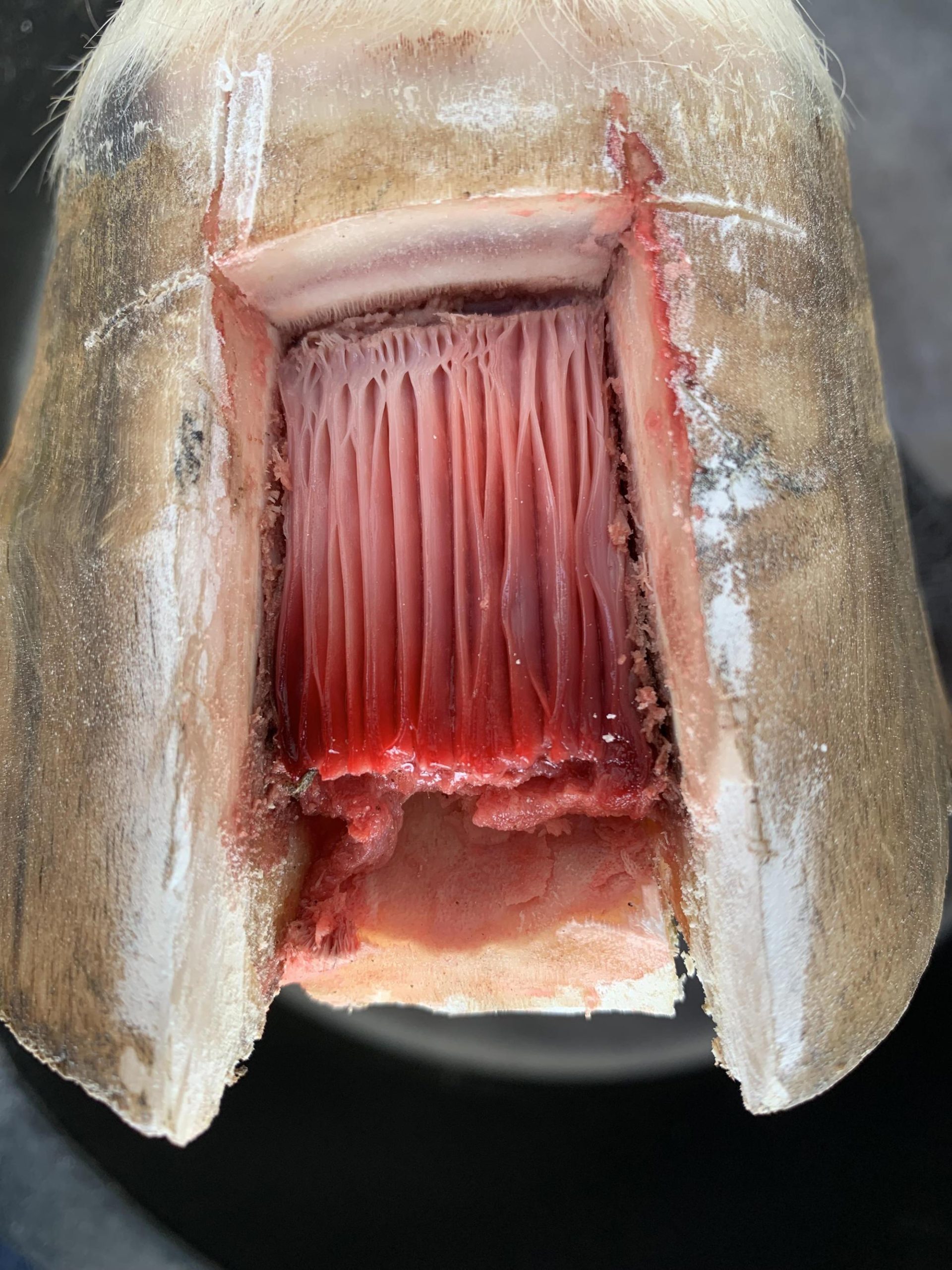
Dermal lamellar
The DDFT, which inserts on the palmar surface of the distal phalanx (P3), exerts a constant tensile force that tends to rotate the toe of P3 downward (“capsular rotation” or “sinking”).
The navicular bone (distal sesamoid) acts as a pulley, redirecting DDFT force.
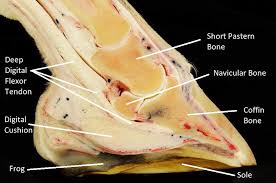
Sagital aspect of the foot highlighting relevant anatomical structures
If the tendon’s angle of pull relative to P3 is steep, tension increases → more force is transmitted dorsally to the lamellae.
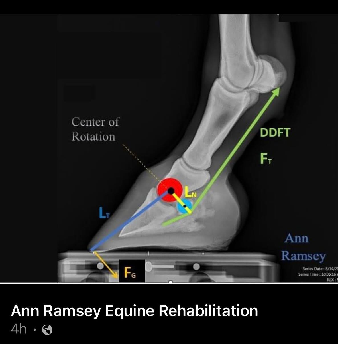
The angle
Of inclination of the DDFT over the DS, and the angle formed between CoR and CoRDs is thought to be a determining factor in tensile force of the DDFT
Effect of heel elevation and palmar extension
Heel elevation raises the palmar aspect of the hoof capsule, increasing the hoof–pastern axis.
This changes the angle of inclination of the DDFT where it passes over the navicular bone.
A more acute hoof-pastern angle reduces the vertical component of DDFT force acting to rotate P3, effectively lowering lamellar strain.
The relationship is trigonometric:
FDDFT,rotational=FDDFT,total⋅sin(θ)
Where:
FDDFT,total = tendon tension (a function of horse mass, gait phase, muscle activity)
θ = angle between DDFT and palmar cortex of P3
Raising the heels reduces θ, therefore reducing the rotational component of tendon force.
The mechanical relationship relieves venous congestion increasing perfusion though both laminar and solar plexus however perfusion is modestly reduced in the loading heel areas
Palmar extension (e.g. extended shoe branch, wedge pad with lever) shifts the ground reaction force (GRF) palmarly.
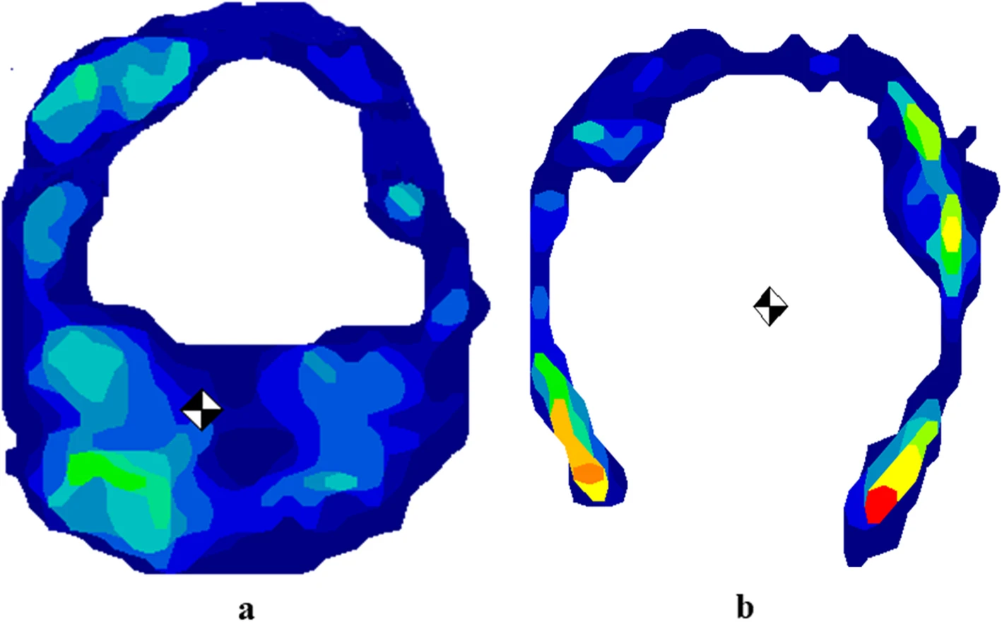
The averaged pressure distribution with the centre of the force ( ) of a horse (number 6) with (a) and without the HCHW (b) during the stance phase. Lateral is to the left and dorsal is in the top. The pressure distribution is colour coded: red = highest pressure forces and deep blue = lowest pressure forces, as seen on the pressure scale. With HCHW, the centre of the force was located palmary in the heel region. The hoof contact area decreased after removal of the HCHW in the heel region and the centre of the force shifted dorsally to the middle hoof region compared with HCHW. BFC = barefoot condition; HCHW = hoof cast with heel elevation
This moves the center of pressure (CoP) closer to the navicular region.
The moment arm between the dorsal lamellae (toe) and the GRF shortens → reducing torque applied, and shortening break over time, to the weakened lamellar system.
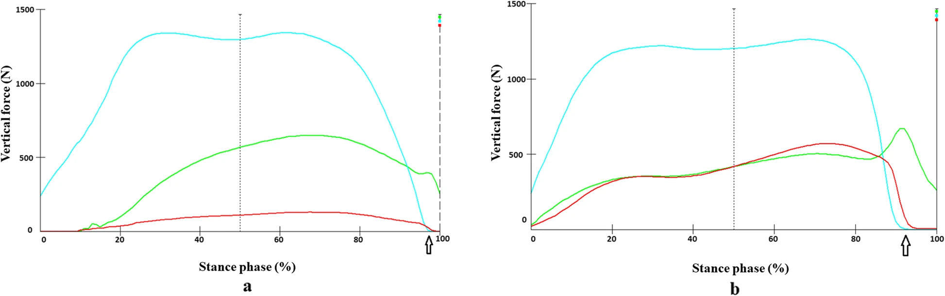
Force-time-curve of the three hoof regions in a representative horse (number 6) with (a) and without an HCHW (b) during stance phase. The heel region (aqua) experienced the highest load in both conditions. The load distributions in the toe (green) and middle (red) regions of the hoof were similar in the BFC. The load in the middle hoof region (red) increased after removal of the HCHW. The arrow indicates the time at which the heel off occurred. BFC = barefoot condition; HCHW = hoof cast with heel elevation
Central loading through the foot
The goal is to centralize GRF through the bony column rather than at the toe.
Mathematically, if:
M=F⋅d = torque about the lamellae
F = ground reaction force (approximated as horse weight / # limbs in stance)
d = horizontal distance between CoP and center of rotation of P3
Then:
Increasing palmar extension reduces d, decreasing torque.
Elevation and wedge design alter force vectors so that GRF is borne more centrally under the distal interphalangeal (DIP) joint.
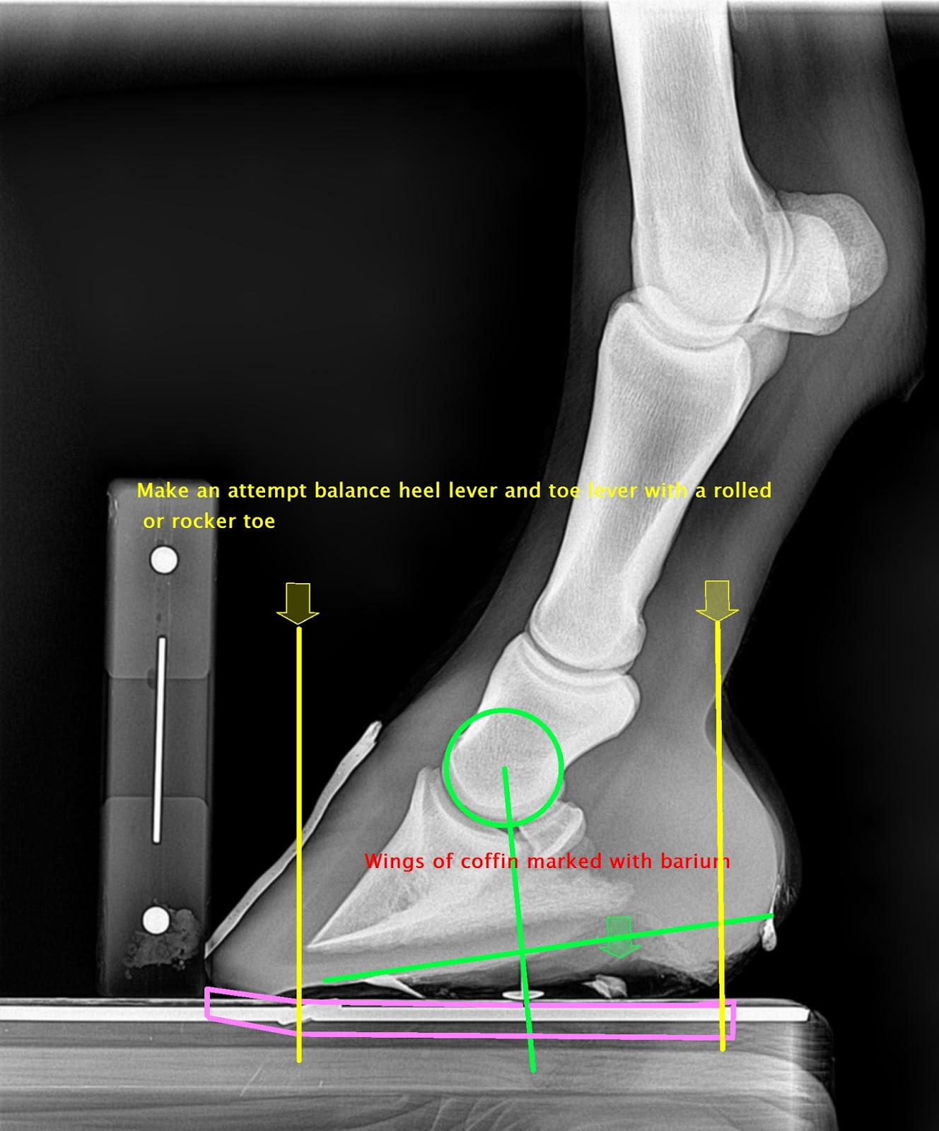
Note the centre if rotation is drawn perpendicular to the bearing surface of the hoof and not the shoe
Shoe length vs. degrees of elevation
There’s a geometric trade-off:
A short wedge (steep angle) concentrates load → unstable and high peak pressures.
A longer palmar extension distributes load over greater surface, allowing lower angle per unit length.
If wedge length = L, elevation height = h, the shoe’s palmar extension angle is:
α=arctan(hL)
For biomechanical efficiency:
You want α sufficient to reduce DDFT torque (usually 6–18° heel lift),
While keeping L long enough to spread load caudally, maintaining central pressure under the DIP joint rather than toe or extreme heel.
A palmar shift in the orientation of CoP as a result of increased elevation is compensated for by an increase palmar bearing border length and rolled toe mechanics
Impact on the digital suspensory system (lamellae)
The lamellae function as a suspensory sling, holding P3 inside the capsule.
Without intervention: dorsal lamellae resist torque of DDFT → tensile overload → separation/failure.
With elevation/extension:
DDFT moment arm shortened, reducing dorsal pull.
CoP shifted palmarly, reducing lever arm.
GRF redistributed centrally → load is shared by solar corium and heel region rather than dorsal lamellae.
Thus, the digital suspensory system is unloaded, slowing progression of mechanical failure in laminitis.
In summary:
Heel elevation reduces DDFT rotational force via trigonometric reduction of tendon angle.
Palmar extension centralizes loading by shifting CoP and reducing torque on lamellae.
The math is essentially about balancing force vectors, torque arms, and pressure distribution:
Frot=FDDFT⋅sin(θ)
M=F⋅d
α=arctan(h/L)
Together, these modifications reduce destructive stresses on the lamellar apparatus while maintaining biomechanical efficiency.
The mechanical relationship relieves venous congestion increasing perfusion though both laminar and solar plexus however perfusion is modestly reduced in the loading heel areas