
The Equine Foot: A Miracle of Bioengineering
Hemodynamic Shock Absorption During the Stance Phase of Locomotion
Caldwell. M. N., Madden. N., & Prout.A. HoofFlix.Com. Email info@hoofflix.com
Introduction
The equine foot exemplifies masterful biomechanical design, combining anatomical structures and fluid dynamics to absorb and dissipate enormous impact forces during locomotion. Rather than acting as a passive structure, the hoof functions as an active hydraulic and viscoelastic damping system, protecting the distal limb from potentially injurious loads. This review synthesises current research on the hemodynamics and mechanical behavior of the hoof throughout the stance phase, focusing on peer-reviewed studies and the physical principles that underpin them.
Hemodynamics and Shock Absorption: The Two-Phase Model
Contemporary understanding frames the hoof’s hydraulic mechanism as operating in two synergistic phases. During the swing phase, arterial inflow predominates—lifted off the ground, hoof tissues engorge with blood while venous valves prevent backflow. Upon hoof impact, the foot transitions into a hydraulic “pump”: blood is forced from the venous plexuses by tissue compression, providing fluid-mediated shock absorption (Bartek Equine LLC, 2024)
Dynamics
As the hoof makes ground contact, impact forces act to decelerate the hoof. Gustås et al. (2006) recorded peak hoof decelerations that are narrow, high-amplitude events consistent with a spring-damper system, where viscoelastic tissues dampen the collision energy PubMed+1ResearchGate+1. These early impulses do not fully decelerate hoof motion—as on sand surfaces—but repeat consistently on firm surfaces, underscoring the role of internal damping elements (Gustås et al., 2006) PubMed. Concurrently, the expanding hoof capsule compresses blood from the venous plexus, a process governed by Poiseuille’s law: small vessel radius amplifies flow resistance, so rapid pressure displacement absorbs energy (Bowker, 2001).
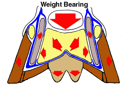
Figure 1. Diagram of hoof expansion and blood displacement during mid‑stance. As the hoof spreads laterally and the digital cushion compresses, venous plexus fluid is pumped upward, converting mechanical shock into hydraulic flow.
Support and Braking Phase: Mid‑Stance to Breakover
After impact, the hoof continues to decelerate longitudinally while in full contact with the ground, a process termed the longitudinal braking phase. Chateau et al. (2010) observed that this phase lasted upwards of 30–40 ms and involved forward sliding of the hoof before stabilisation, accompanied by oscillatory vibrations from internal structuresPubMed+5Beva Online Library+5PubMed+5PubMed+2Wikipedia+2. Pressure sensors recorded a drop in digital cushion pressure during this phase, indicating tissue compression rather than active loading, challenging theories that load is supported by frog weight alone (Van Heel et al., 2010) Beva Online Library.
Material Properties: Viscoelasticity and Hydration Effects
Finite element modelling reveals that hoof capsule mechanics depend heavily on its hydration and material composition. A hyperelastic Mooney–Rivlin model (Marlow formulation) better replicates true stress–strain behavior compared to linear elastic approximations (Akbari Shahkhosravi et al., 2021) PubMed+1Wiley Online Library+1. Increased hydration results in lower tissue stiffness and higher strain under load, indicating potential seasonal or environmental impact on risk of injury.
Anatomical Pathways for Fluid Return
The hoof’s venous drainage system is essential for hydraulic pumping. Van Heel et al. (2010) described the hoof wall and digital cushion draining via the coronary venous plexus, inner corium plexus, and heel tributaries, most of which are valveless—facilitating efficient fluid removal during stance loading Wikipedia+3ResearchGate+3Wikipedia+3. Efficient venous return is foundational to the hoof’s shock absorption function.
Physical Principles Applied
Hooke’s Law: Elastic deformation of hoof wall and soft tissues absorbs instantaneous force.
Poiseuille’s Law: Resistance to blood flow central to hydraulic damping during arterial-to-venous displacement.
Impulse–Momentum Principle: Extending force duration lowers peak internal load.
Conservation of Energy: Transformation of kinetic energy into fluid flow and viscoelastic deformation dissipates impact force.
Consequences of Conformation or Shoeing Alterations
Deviations from optimal hoof structure impair the shock‑absorbing system. Shoeing traditional steel shoes restrict heel expansion by up to 36%, thereby reducing both hydraulic function and hoof-pastern axis dynamics (Bartek Equine LLC, 2024) 1ResearchGate+1Bartek Equine LLC+1PubMed+1. Reduced hemodyamic compliance increases transmitted vibration and peak accelerations to the first phalanx (Van Heel et al., 2010) PubMed. Excessive toe length, underrun heels, or contracted frogs further reduce ability for fascia and dermal stretching and frog-ground contact, leading to peripheral loading and increased risk of laminitis or navicular degeneration.
Clinical Implications for Farriery and Rehabilitation
Effective trimming and shoeing should aim to preserve physiological hoof expansion and vascular return. Ideal interventions include trimming to restore dorsopalmar alignment, ensuring frog engagement, and using shoe designs that permit heel movement—such as rocker toes, wide-webbed shoes, or impact-absorbing pads. These approaches support the hoof’s natural damping capabilities, in line with biomechanical and haemodynamic principles, thereby reducing risk of foot pathologies.
Conclusion
The equine foot represents a biologically engineered marvel, combining mechanical design and fluid dynamics to manage extreme forces encountered during locomotion. Understanding the synchronized roles of vascular pumping, tissue viscoelasticity, and structural compliance provides essential insight into preventive and rehabilitative hoof care. Ongoing research into hoof material properties and fluid dynamics promises further enhancement of farriery science and equine welfare.
for more information watch our video tutorial at https://hoofflix.com/media/videos/anatomy-of-lower-limb-and-foot/vascular-supply-to-the-equine-limb/
References
Akbari Shahkhosravi, N. et al. (2021). Finite element modeling of equine hoof biomechanics using hyperelastic material models. Journal of Biomechanics, 128:110715 PubMed.
Bartek Equine LLC. (2024). Blood Circulation in the Hoof. Bartek Equine Education Bartek Equine LLC.
Chateau, H. et al. (2010). Biomechanical hoof-ground interaction on different surfaces. Equine Veterinary Journal, 42(3):247–253 ResearchGate+7Beva Online Library+7PubMed+7.
Denoix, J.-M. (2019). Diagnosis and Management of Lameness in the Horse. 2nd ed., Saunders Elsevier.
Gustås, P. et al. (2006). Time-domain characteristics of hoof-ground interaction. Equine Veterinary Journal, 38(7):634–641 Wikipedia+2PubMed+2ResearchGate+2.
Murray, R. C. et al. (2006). MRI investigation of foot lameness. Equine Veterinary Education, 18(6):335–342.
O’Grady, S. E. & Poupard, D. A. (2003). Heel pain foot care techniques. Veterinary Clinics of North America: Equine Practice, 19:365–385.
Schramme, M. C. et al. (2020). Diagnostic imaging of the equine foot. Veterinary Radiology & Ultrasound, 61(4):393–405.
Van Heel, M. C. V. et al. (2010). Effect of trimming on hoof balance via dynamic pressure measurement. Equine Veterinary Journal, 36(8):778–782 Beva Online Library.