
Suspensory Ligament Desmopathy in the Horse:
Biomechanical Pathophysiology, Diagnostic Integration and the Central Role of Therapeutic Farriery in Rehabilitation
Mark Caldwell. PhD., FWCF. HoofFlix at Scientific Horseshoeing.
Abstract
Suspensory ligament desmopathy (SLD) remains one of the most prevalent performance-limiting conditions in sport horses and racehorses. While advances in diagnostic imaging and regenerative therapeutics have refined clinical management strategies, the biomechanical environment in which the suspensory apparatus functions remains the most significant determinant of both lesion development and successful rehabilitation. The suspensory ligament is a passive energy-storage structure that is subjected to substantial cyclic strain during locomotion, particularly during mid-stance, when fetlock hyperextension is maximal. Alterations in hoof balance, toe length, heel height and mediolateral symmetry directly influence the magnitude and duration of suspensory strain.
This article synthesises current scientific understanding of suspensory biomechanics, clinical diagnosis and veterinary treatment options, with particular emphasis on the evidence-based role of therapeutic trimming and shoeing. Farriery interventions are examined not merely as adjunctive measures, but as primary biomechanical strategies that modulate strain distribution, reduce pathological loading and facilitate structured rehabilitation.
Introduction
Suspensory ligament desmopathy encompasses acute fibre disruption, chronic degenerative change, proximal suspensory desmitis (PSD), branch injuries and insertional pathology. The condition affects both forelimbs and hindlimbs, although hindlimb proximal PSD is increasingly recognised as a distinct clinical syndrome in sport horses (Dyson, 2011).
Despite advances in regenerative therapies, recurrence rates remain significant. This highlights the need to address mechanical loading factors intrinsic to hoof conformation and farriery practice.
The suspensory ligament does not fail in isolation; it fails within a biomechanical system.
Functional Anatomy and Biomechanics
The suspensory ligament (interosseous medius) originates from the proximopalmar or proximoplantar aspect of the third metacarpal/metatarsal bone and adjacent carpal/tarsal bones. It divides distally into medial and lateral branches inserting on the abaxial surfaces of the proximal sesamoid bones, with extensor branches contributing to the common or long digital extensor tendon (Denoix, 1994; Denoix, 2014).
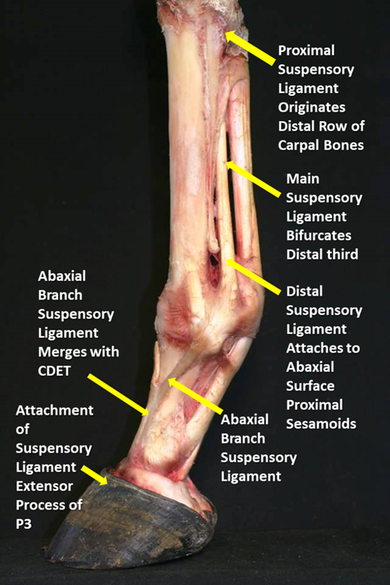
The origin, route and insertions of the suspensory ligament: photo credit Wayne Preece FWCF
It functions as part of the suspensory apparatus, resisting fetlock hyperextension and storing elastic strain energy during locomotion.
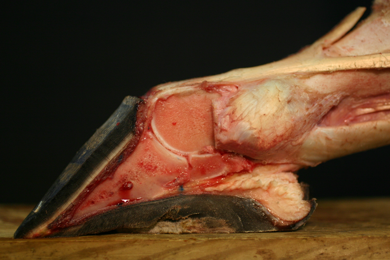
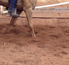
Dorso flexion: photo credit Mitch Taylor
Strain During Locomotion
Peak suspensory strain occurs during mid-stance when vertical ground reaction force (GRF) is maximal and the fetlock joint reaches its greatest degree of extension (Smith et al., 2006; Clayton and Hobbs, 2017).
In galloping Thoroughbreds, strain values approach 12–16%, near physiological tolerance limits (Smith et al., 2006). Cumulative cyclic loading is therefore central to pathogenesis.
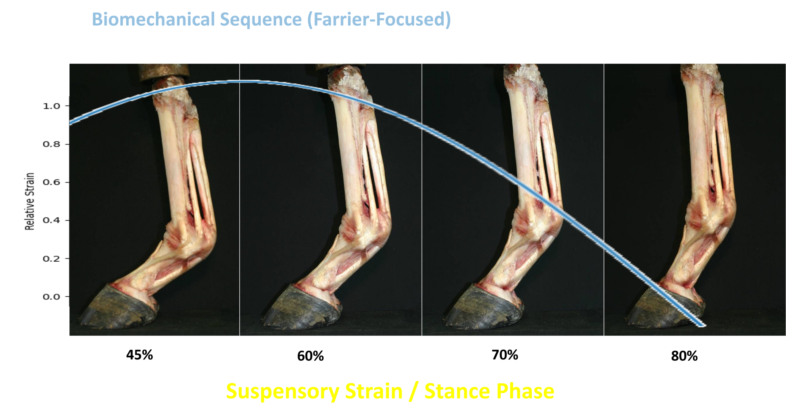
peak force time line of the suspensory ligament from maximum at mid stance to breakover.
Pathophysiology
Acute lesions involve fibre disruption and haemorrhage. Chronic cases demonstrate collagen disorganisation, neovascularisation and fibrosis. Chronic hindlimb PSD frequently demonstrates concurrent bone oedema and enthesopathy detectable via MRI (Dyson, 2011).
Collagen Architecture
Normal ligament demonstrates highly aligned Type I collagen fibres. Chronic desmopathy results in disorganised fibre orientation and inferior mechanical properties.
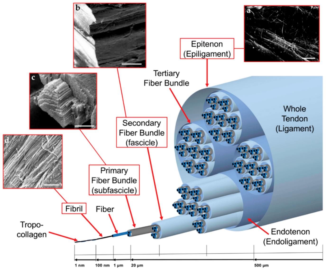
Architecture of the suspensory ligament. Biofabrication of electrospun scaffolds for the regeneration of tendons and ligaments (credit Sensini A and Cristofolini L 2018).
Diagnostic Integration
Clinical Signs
Insidious lameness
Reduced impulsion (hindlimb PSD)
Pain on proximal palpation
Reduced fetlock extension
Diagnostic Analgesia
Regional analgesia including high 4-point blocks and proximal metacarpal/metatarsal blocks assists localisation. Care must be taken to differentiate from DDFT analgesia.
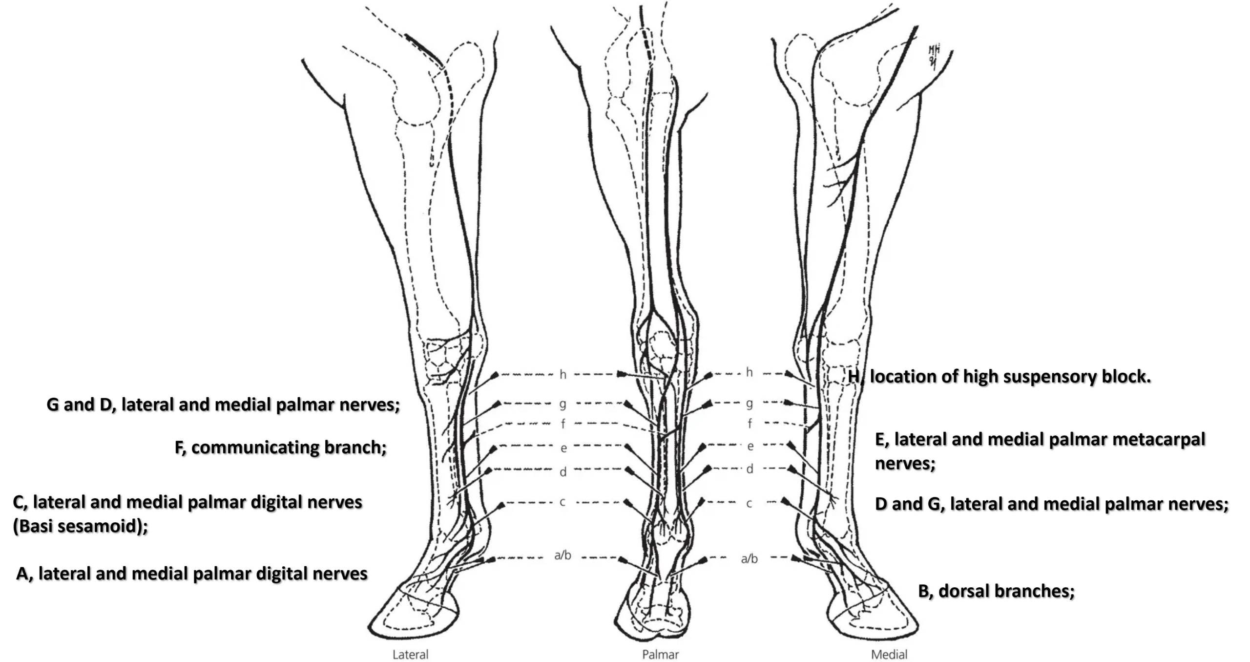
Regional analgesia including high 4-point blocks and proximal metacarpal/metatarsal blocks assists localisation.
Diagnostic Imaging
Ultrasonography remains first-line for branch injuries. MRI is considered gold standard for hindlimb PSD, particularly where bone involvement is suspected (Dyson, 2011).
Transverse image of a proximal suspensory ligament grade 1 lesion about 4 cm distal to the head of the MTII. The lesion appears as a focal, well-defined, hypoechogenic area (white arrows) in the ligament occupying <25% of the cross-sectional areal. Longitudinally the lesion is well-demarcated (white arrows), it begins from the insertion and extends distally. In transverse image medial is to the left; in longitudinal image proximal is to the left. Kendra D. Freeman et al 2025
Veterinary Treatment Modalities
Controlled exercise remains fundamental, promoting fibre alignment during repair (Smith et al., 2006).
Extracorporeal shockwave therapy demonstrates moderate benefit in proximal PSD (Dyson, 2011).
Surgical plantar fasciotomy and neurectomy have reported return-to-work rates of 60–80% in selected hindlimb PSD cases (O’Meara et al., 2012).
However, none of these interventions modify the primary biomechanical loading environment. This responsibility lies with farriery.
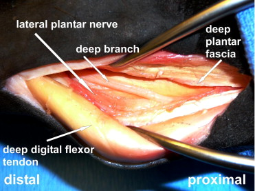
Neurectomy of the deep penetrating branch of the lateral plantar nerve at the level of the proximal metatarsus is used to reduce pain and swelling associated with suspensory ligament desmitis. The technique is most applicable to chronic, poorly healing suspensory desmitis or recurrent desmitis.
The Biomechanical Basis for Therapeutic Farriery
The magnitude and duration of suspensory strain are influenced by:
Toe length (moment arm)
Heel height and base of support
Medioloateral balance
Surface interaction
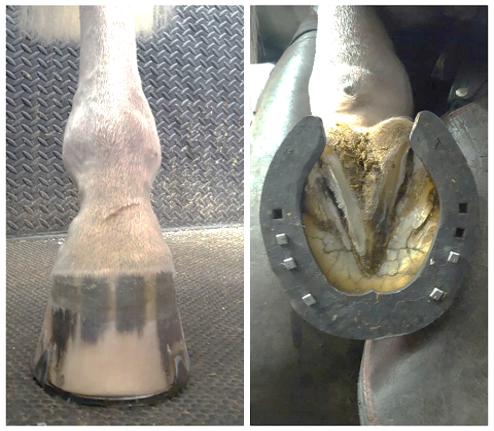
Toe Length and Lever Arm Mechanics
A longer toe increases the digital flexor moment arm, prolonging breakover and increasing the duration of peak fetlock extension (Clayton and Hobbs, 2017). Therapeutic trimming should therefore prioritise physiological toe shortening to reduce lever arm effects without excessive dorsal wall thinning.
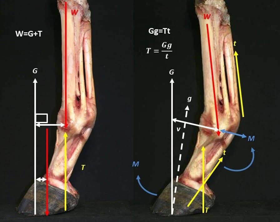
The extensor moment acting on the limb is calculated by the ground reaction force acting through the COP times the distance of the COP from the centres of rotation. The extensor moment is a rotational and collapsing force acting on the limb.
Shoeing Modalities in Suspensory Rehabilitation
Reframing the Problem: The Suspensory Ligament as a Time-Dependent Strain Structure
The suspensory ligament is not injured purely by peak load. It is injured by:
Magnitude of peak strain
Duration of strain
Rate of strain application
Number of loading cycles
Farriery interventions can modify:
Breakover timing
Centre of pressure (COP) migration
Fetlock extension amplitude
Torsional loading at branch insertions
Shear forces generated by surface interaction
The complexity arises because shoe effects are surface-dependent, and certain interventions may reduce peak strain but increase cumulative strain duration — a critical and often overlooked issue.
Breakover Mechanics and Shoe Type Selection – The Digital Flexor Moment Arm
Toe length increases the moment arm acting around the distal interphalangeal joint. This delays heel lift and prolongs fetlock hyperextension, increasing suspensory strain duration (Clayton and Hobbs, 2017).
However, excessive reduction of dorsal wall length without regard to hoof capsule morphology may destabilise distal phalanx alignment.
Rolled Toe and Rocker Toe Shoes – Biomechanical Effects
Earlier breakover
Reduced stance duration
Reduced duration of peak fetlock extension
Potential reduction in cumulative strain
Counterintuitive Consideration
On deep surfaces, earlier breakover may:
Increase vertical impulse earlier in stance
Increase muscular demand to stabilise the limb
Increase fatigue-related cumulative strain
Thus, a rocker toe may reduce peak strain on firm ground but increase fatigue loading in deep arena footing.
Denoix (1999, 2014) emphasised that distal limb mechanical adjustments influence proximal limb strain patterns. Excessively aggressive rocker profiles may also increase instability during early stance, especially in horses with hindlimb PSD.
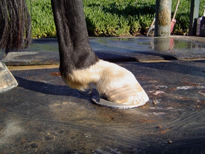
A roller motion / rocker toe shoe may be counterintuative on artificial arena surfaces. Photo credit Peter Peers DWCF
Square Toe Shoes
Square toes reduce breakover lever arm in the sagittal plane but may increase mediolateral instability if not correctly aligned with the limb axis.
In horses with branch lesions, especially unilateral medial branch injury, a square toe may increase torsional asymmetry unless carefully balanced.
Heel Support Modalities – Egg Bar Shoes
Intended Effect
Increase palmar/plantar ground contact
Reduce fetlock hyperextension amplitude
Shift centre of pressure caudally
Surface Dependent Consideration
On firm surfaces:
Increased caudal support may reduce hyperextension.
On deep surfaces:
Increased surface engagement may increase drag.
Increased drag increases muscular effort and cumulative strain.
Counterintuitively, egg bars on deep arena surfaces may increase fatigue loading and exacerbate chronic hindlimb PSD.
Straight Bar Shoes
Straight bars provide mediolateral stability and may reduce branch torsion. However:
On uneven terrain, increased surface area may increase asymmetric shear forces.
Excessive rigidity may reduce adaptive hoof deformation.
Suspensory branches are particularly sensitive to torsional loading (Denoix, 2014).
Counterintuitively, egg bars on deep arena surfaces may increase fatigue loading and exacerbate chronic hindlimb PSD.
Heel Elevation: A Double-Edged Intervention – Heel wedges are sometimes used in proximal suspensory pathology.
Proposed Rationale
Reduce DDFT tension
Reduce fetlock extension moment
However, biomechanical modelling suggests that excessive heel elevation:
Increases compressive force within distal interphalangeal joint
May increase strain on distal sesamoidean ligaments
Alters proximal limb kinematics unpredictably
Clayton and Hobbs (2017) demonstrated that small angular changes in hoof orientation significantly alter proximal limb loading.
For hindlimb PSD, Denoix (2014) cautioned against excessive elevation, particularly in horses with straight hock conformation.
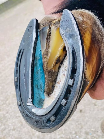
Heel elevation increases dorsiflexion of the fetlock and is contra indicated for suspensory issues.
Wide-Web Shoes
Wide-web designs increase load distribution across the solar surface.
Benefit:
Reduce peak pressure
Reduce focal overload
Limitation:
On soft surfaces, wider web increases suction effect and resistance during breakover, potentially increasing stance duration.
This demonstrates the time-dependent paradox: Reduced peak force may coexist with increased time under load.
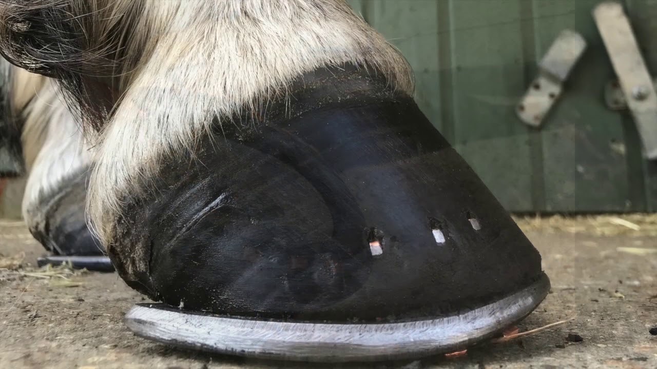
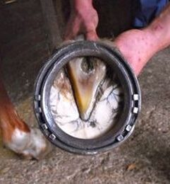
Selective additional width of section reduces peak pressure by increasing the ground surface area. It is thought to be most effective when selection and application are anatomically sympathetic to the lession.
Surface Interaction: The Overlooked Variable
Surface characteristics modify:
Vertical GRF magnitude
Rate of loading
Shear forces
Limb deceleration forces
Fatigue accumulation
(Murray et al., 2010; Clayton and Hobbs, 2017)
Firm Surfaces
Characteristics:
Higher peak GRF
Shorter stance time
Lower fatigue accumulation
Implication:
Shoes that reduce peak strain (rolled toe, moderate heel support) are often effective.
Deep Surfaces
Characteristics:
Lower peak GRF
Longer stance duration
Increased muscular effort
Increased fatigue cycles
Implication:
Interventions that increase ground engagement (wide-web, large bars) may increase drag and cumulative strain.
Counterintuitively:
A more minimal shoe with optimised breakover may be preferable to heavy supportive bar shoes in horses working primarily on deep surfaces.
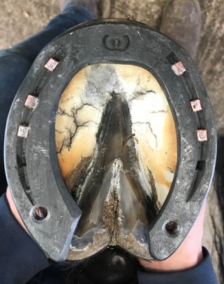
Selection and application of therapuetic shoes is surface dependent.
Synthetic Arena Surfaces
High shear forces may increase torsional branch loading.
In such cases:
Medioloateral balance becomes critical.
Asymmetrical breakover can exacerbate branch strain.
Summary
Comparative Shoeing Strategy by Lesion Type
| Lesion Type | Preferred Intervention | Surface Consideration | Caution |
|---|---|---|---|
| Forelimb Branch | Mild rolled toe + mediolateral precision | Avoid torsion on uneven terrain | Avoid excessive square toe |
| Hindlimb PSD | Moderate toe reduction + conservative heel support | Avoid deep, fatiguing surfaces | Avoid excessive wedges |
| Chronic Degenerative | Conservative breakover + progressive loading | Monitor fatigue accumulation | Avoid aggressive rocker profiles |
| Bilateral PSD | Symmetry paramount | Surface conditioning essential | Avoid asymmetrical shoeing |
Rehabilitation Phasing and Shoeing Progression
Phase 1: Acute
Conservative toe shortening
Moderate caudal support
Firm, level surfaces
Phase 2: Controlled Loading
Maintain optimised breakover
Reduce excessive support gradually
Avoid deep footing
Phase 3: Return to Work
Discipline-specific shoeing
Surface simulation during rehab
Continuous reassessment
Smith et al. (2006) emphasise that progressive mechanical loading promotes organised collagen alignment. Farriery must evolve during healing.
The Counterintuitive Lessons
More support does not always reduce strain.
Increased surface contact may increase fatigue loading.
Peak force reduction is less important than strain duration.
Heel elevation may destabilise proximal biomechanics.
Surface interaction can negate shoeing benefits.
The suspensory ligament responds to mechanical environment over time, not single interventions.
Integrating Denoix’s Functional Framework
Denoix’s work repeatedly demonstrates that:
Distal limb mechanics alter proximal strain distribution.
Hoof balance affects limb axis alignment.
Fetlock kinematics are sensitive to small angular hoof changes.
(Denoix, 1994; Denoix, 1999; Denoix, 2014)
His functional anatomy model supports the concept that therapeutic farriery is a primary biomechanical intervention, not a secondary adjunct.
Evidence-Based Recommendations
Prioritise controlled breakover over aggressive mechanical alteration.
Avoid heavy bar shoes on deep surfaces without justification.
Consider surface as part of prescription.
Reassess shoeing at each rehab stage.
Monitor imaging progression alongside mechanical adjustments.
References
Clayton, H.M. and Hobbs, S.J. (2017) ‘The role of biomechanical analysis in equine locomotion research’, Equine Veterinary Journal, 49(5), pp. 560–568.
Denoix, J.M. (1994) ‘Functional anatomy of the suspensory apparatus’, Equine Veterinary Education, 6(4), pp. 189–197.
Denoix, J.M. (1999) ‘Biomechanical approach to distal limb pathology’, Proceedings of the AAEP, 45, pp. 294–298.
Denoix, J.M. (2014) ‘Functional anatomy and diagnostic imaging of suspensory ligament injuries’, Veterinary Clinics of North America: Equine Practice, 30(1), pp. 29–50.
Murray, R.C., Walters, J.M., Snart, H., Dyson, S.J. and Parkin, T.D.H. (2010) ‘Identification of risk factors for superficial digital flexor tendon injury in National Hunt racehorses’, Equine Veterinary Journal, 42(3), pp. 228–234.
Smith, R.K.W., Birch, H.L., Patterson-Kane, J.C. and Firth, E.C. (2006) ‘The pathophysiology of tendon and ligament injury’, Equine Veterinary Journal, 38(2), pp. 130–136.