
Stop Looking at the Feet First
Why every foot assessment should begin with the whole horse — and why the foot that baffles you is usually explained by something above it
Mark Caldwell PhD., FWCF & Neil Madden FWCF · From: Reading the Whole Horse — The Equine Foot, Vol. I
A ten-year-old Warmblood gelding. Regular shoeing, six-weekly, balanced trim every cycle. And yet the left hind capsule keeps doing the same thing: medial wall thickening, slight lateral flare, a morphology that drifts back within weeks of every re-shoeing. Three farriers have worked on this horse. The third one — before lifting a single foot — asks a question the previous two never asked. Can I see this horse walk, please? And can I look at it from behind first?
What follows changes the clinical picture entirely: the right gluteal muscle is visibly flatter than the left. The tuber sacrale is asymmetric. This horse has been loading its left hind as a compensatory limb for months. The foot is not causing the problem. The foot is recording it.
The Chain, Not the Last Link
The Equine Foot introduces a framework that changes how we think about the foot’s role in the locomotor system. The equine locomotor system is not a collection of anatomical departments that happen to share a skeleton. It is a single integrated mechanical chain — from the cervical vertebrae at the poll, through the thorax and back, through the hindquarter and the forelimb, to the foot at the ground. Every link influences every other link. And the foot is the terminal element: the point where the chain meets the ground, where all proximal variation is expressed, and where the farrier has the most direct, most regular, most mechanically precise access.
This is not a romantic metaphor. It is a measurable mechanical reality. When the thoracolumbar junction at T13 to L3 is compromised by kissing spines or facet joint pathology, the efficiency of hindquarter propulsive force transfer to the forelimbs is reduced. The horse compensates by altering how it loads its feet — weeks before any structural foot change is visible. The farrier who is watching the right things at the right time can see this change in progress.
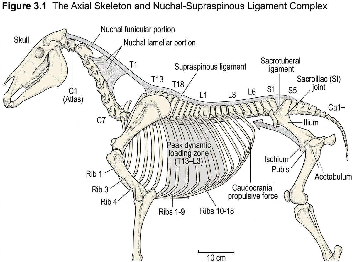
Fig 3.1: Full lateral axial skeleton — nuchal/supraspinous complex, vertebral regions, T13-L3 highlighted, sacroiliac joint and tuber sacrale
“The practitioner who confines their anatomical knowledge to the foot is working with the last few links of a very long chain and ignoring everything that determines how those links are loaded.”
The Muscle You Don’t See
One of the most important anatomical facts about the horse that most farriers never formally learn: the forelimb has no bony articulation with the thorax. No clavicle. No sternoclavicular joint. The entire weight of the trunk — 400 to 600 kilograms — is carried between the forelimbs on a purely muscular and fascial sling. The primary suspensory muscle is serratus ventralis, running from the first eight ribs to the medial face of the scapula.
When serratus ventralis atrophies — from long thoracic nerve damage, chronic thoracolumbar pain, or simple ageing — the scapula wings away from the thoracic wall during loading. The horse compensates by landing harder and faster on the affected foot. That extra loading starts to show up in the capsule within weeks. The persistent toe-first landing in the forelimb that no amount of foot work has resolved? In many of those cases, nobody has looked at the thoracic sling.
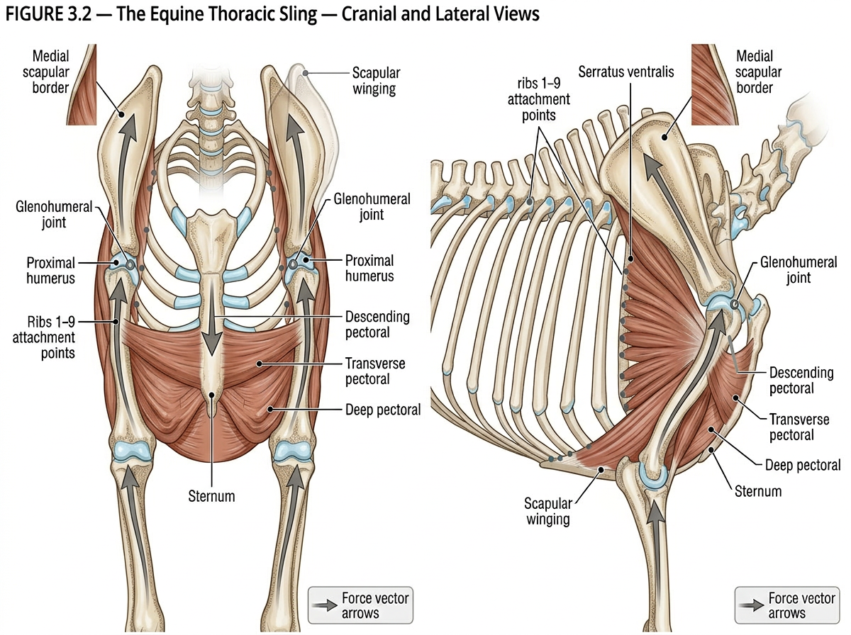
Fig 3.2: Cranial + lateral views of the thoracic sling — serratus ventralis and pectoral group
Reading the Gluteals Before the Feet
The hindquarters generate the majority of net forward propulsion in equine locomotion. The gluteal group drives hip extension in the propulsive phase and forms the visible bulk of the rump. Gluteal atrophy is not a subtle finding when it is established: one side noticeably flatter, lower, less filled out than the other. That asymmetry has been building for weeks or months. A horse does not lose muscle mass from a healthy limb it is using normally.
When gluteal atrophy is contralateral to the more heavily worn or loaded hind foot, the clinical picture is a compensatory loading shift: the horse protecting the atrophied side by overloading the other. This is one of the most clinically useful observations a farrier can make before lifting a single foot. It tells you which foot is bearing the load, which foot is being protected, and why the overloaded foot looks the way it does.
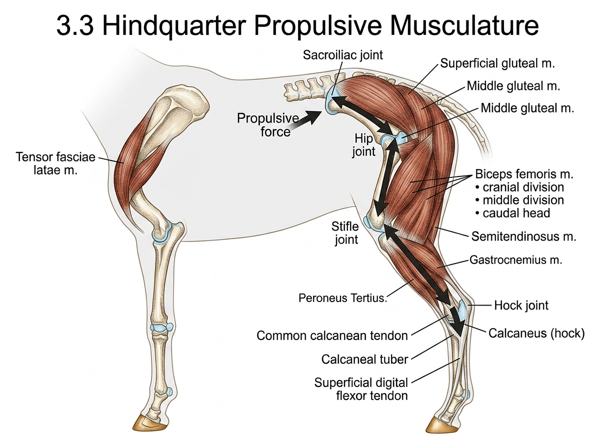
Fig 3.3: Hindquarter propulsive muscular chain — gluteals, hamstrings, gastrocnemius + SDF, calcaneal insertion
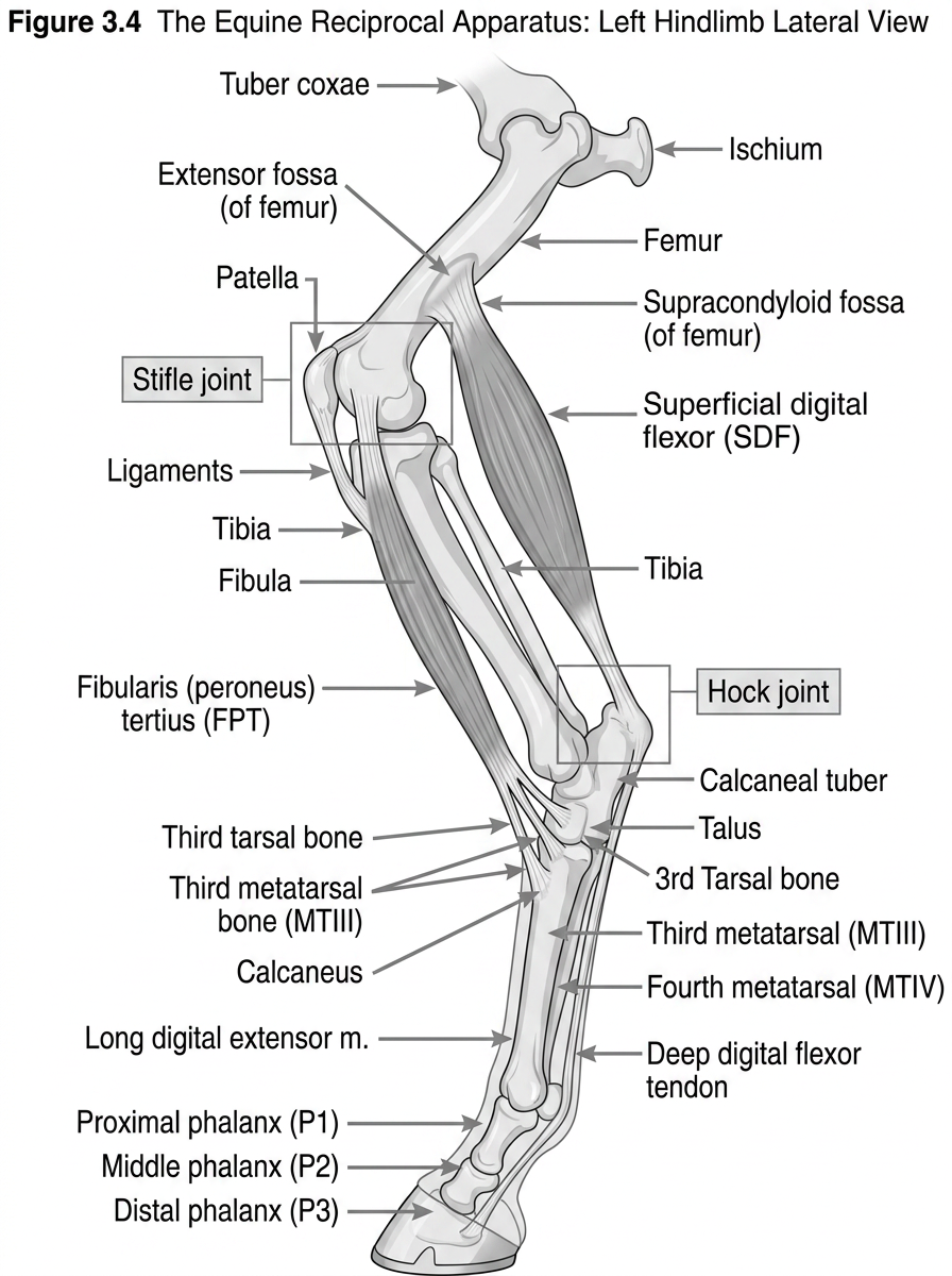
Fig 3.4: Reciprocal apparatus — fibularis tertius and SDF components, showing obligate stifle-hock coupling
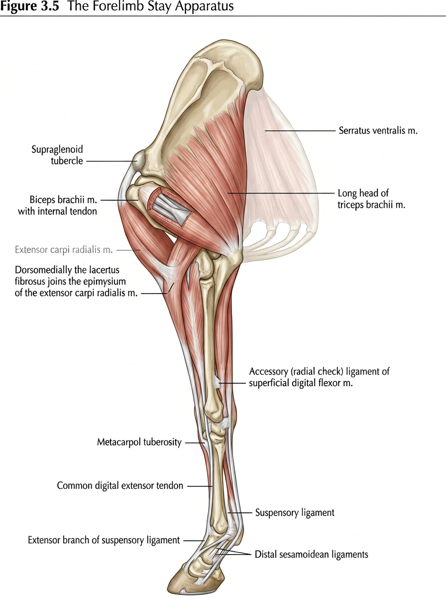
Fig 3.5: Forelimb passive stay apparatus chain — coffin joint to shoulder (DDFT, check ligaments, suspensory, biceps-lacertus)
When Geometry Is Destiny
Conformation is not about aesthetics. In the context of farriery, conformation is a force vector description. Every degree of deviation from the classical ideal is a loading vector that departs from the vertical, applied at every joint below the deviation, at every stride, for the horse’s entire working life. The foot is downstream of everything and cannot escape what is happening above it.
A base-wide horse will overload the medial wall. Every time. Until it is shod to redistribute load, or the proximal geometry is addressed, or the horse retires. The capsule changes are predictable, directional, and mechanical. When a farrier trims that foot to correct the capsule asymmetry and it returns within the shoeing cycle, it returns because the conformation is still generating the loading pattern that produced the asymmetry in the first place.
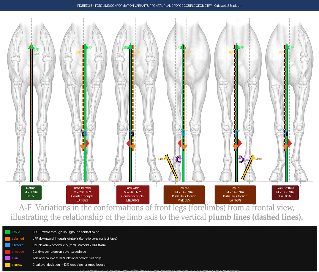
Fig 3.6: Conformational deviations and foot capsule consequences — forelimb (base-wide, base-narrow, toe-in, toe-out) and hindlimb (sickle, straight, cow hock) with force vector overlays and resulting capsule morphology
| The Synthesis Point The foot, read in the context of the whole horse’s conformation, is a structural biography of its mechanical history. The farrier who looks at the horse before looking at the feet provides a quality of assessment that mechanical skill alone cannot replicate. |
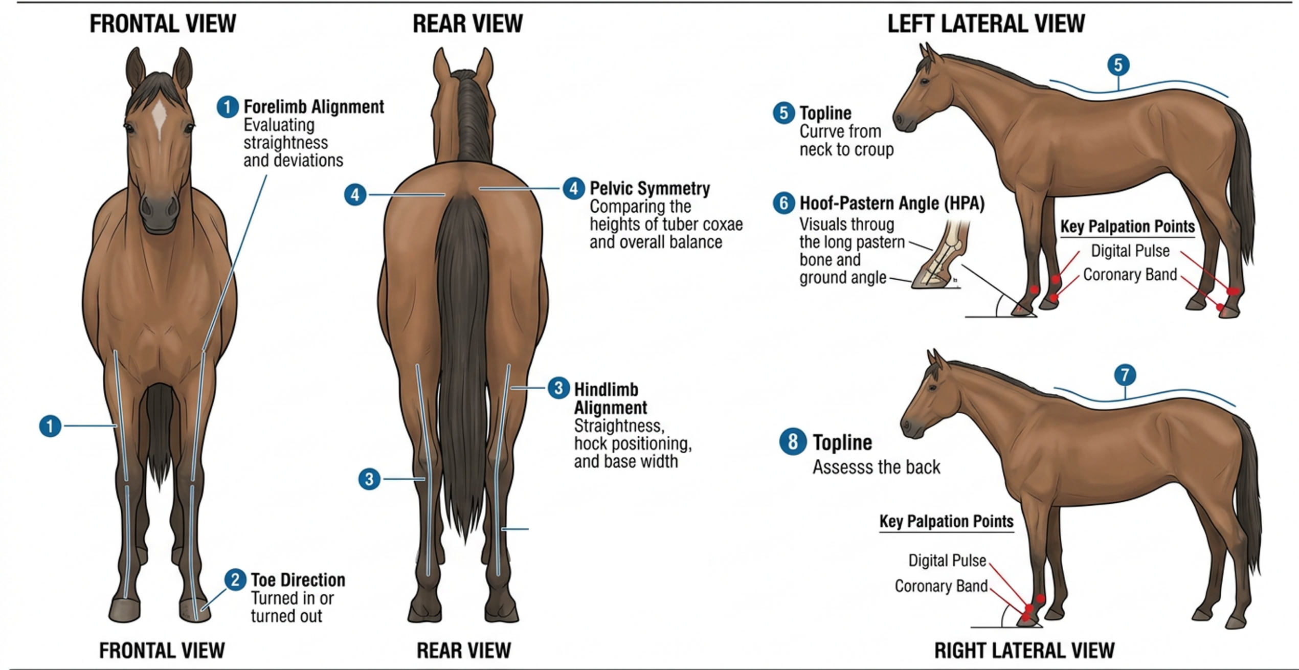
Fig; 3.7 The Four Point Standing Assessment.
What This Article Is (and Is Not)
This is not a comprehensive anatomy text. It is the integrating framework — the functional logic that connects what happens at the poll to what the farrier finds at the foot six weeks later. The practical take-home is Table 3.1, the Regional Reference Summary, which maps each locomotor region to its primary functional role and the clinical signal it generates at the foot when compromised.
Use it as a pre-shoeing mental checklist. Before you pick up a foot, run these rows. Where do you see asymmetry? That row tells you what to expect at the foot. When the foot matches the prediction, you have found a proximal driver. When it does not, that is your cue to investigate the foot itself as the primary site. This is clinical reasoning applied to routine farriery.
Tags: #TheEquineFoot #WholeHorseAssessment #ILSmodel #FarrieryCPD #EvidenceBasedFarriery #ScientificHorseshoeing #HoofFlix #ClinicalReasoning #EquineAnatomy
Mark N. Caldwell PhD FWCF & Neil Madden FWCF · The Equine Foot, Volume I · Scientific Horseshoeing Limited / HoofFlix TV
The Equine Foot: Science, Craft & Clinical Reasoning in Farriery and Podiatry Caldwell & Madden · Scientific Horseshoeing Limited / HoofFlix TV · © 2025