
Sidebone in the Horse: Beyond the Quiet Ossification
Dr. Mark N. Caldwell PhD., F.W.C.F. HoofFlix.CoM
Many horse owners, riders, trainers or farriers have heard of sidebone—the ossification of the collateral (ungual) cartilages of the coffin bone. Often it is regarded as an incidental radiographic finding, something to note, but not something that truly affects performance. But scratch the surface, especially in high-level athletes such as dressage horses, and you find a more complex story—one of subtle loss of soundness, performance compromise, and a potential for prevention and improvement through skilled veterinary and farrier intervention.
This essay seeks to synthesize what is known about sidebone: its causes, its anatomical relationships, how it shows up clinically, how we test for it, and importantly, how farriers (with veterinarians) can manage it with more traditional, refined shoeing methods that go beyond generic bar shoes or wedges. I want to convince you this is a pathology worth watching, diagnosing properly, and treating with finesse.
Anatomy, Pathology, and Why Sidebone Isn’t Always Innocent
The collateral cartilages are paired structures, one medial and one lateral, attached to the lower (palmar or plantar) margins of the distal phalanx. They help the hoof capsule expand and contract under load, dampening concussion, absorbing impact, and modulating hoof capsule flexibility. They are intimately related to:
- The coffin joint (distal interphalangeal joint), including its capsule and synovial lining;
- The insertion and sheath of the deep digital flexor tendon (DDFT) and its navicular apparatus;
- The soft tissues of the heels, including the digital cushion and heel bulbs;
- The hoof capsule itself, which must flex, expand and contract, especially at the heels.
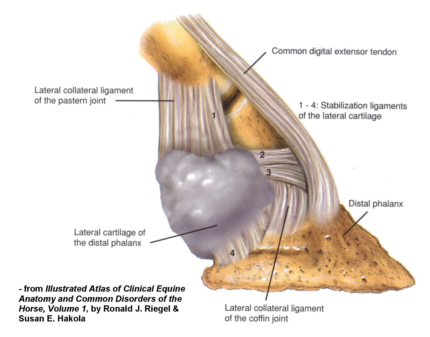
Fig, 1 A schematic: lateral and cross-sectional view of hoof; show collateral cartilages (medial/lateral), distal phalanx, coffin joint, DDFT, navicular bone, digital cushion. Annotate where ossification occurs;
In sidebone, cartilage undergoes ossification via endochondral ossification—microtrauma or chronic concussion to the cartilages leads to chondroid degeneration, mineralization, and sometimes formation of separate ossification centers. Over time an ossified mass may grow proximally, or occasionally fracture, and may impinge upon or interfere with joint spaces or adjacent soft tissues. Even if the ossification is stable, the loss of flexibility at the heel can alter impact absorption, circulation, hoof expansion, and mechanics of breakover.
In practice, this means even horses “with sidebone, but no lameness” may display residual loss of performance: less impulsion, shorter stride, stiffness, slower recovery after hard work, sensitivity on hard ground, reduced ability to execute fine dressage movements that depend on precise weight transfer and lateral balance.
Clinical Signs, Diagnostics, and Prognosis
Clinically, signs may be subtle. Riders might notice:
- Unevenness in transition, especially into lateral or collected work.
- Discomfort on hard, irregular or concussive surfaces.
- Tenderness at the heel area on palpation of collateral cartilage (sometimes).
- Lameness that worsens with tiring or on circles, but improves with soft footing or rest.
- Changes in hoof shape over time—heels may become contracted, distortion of hoof capsule medially or laterally, reduced heel expansion.
- Irregular shoe wear patterns.
Diagnostic approaches include:
- Palpation of collateral cartilages, comparison between sides, checking for heat, swelling, sensitivity.
- Gait analysis, including straight lines, circles, different surfaces.
- Diagnostic analgesia (nerve/joint blocks) to localise pain: palmar digital blocks, coffin joint blocks.
- Radiography to see the ossified cartilage, its size, orientation, whether there are fractures or lucent lines, relationship with the distal phalanx or coffin joint.
- Advanced imaging (ultrasound, MRI, CT) in higher-value horses or where radiographs don’t explain all of the lameness. MRI can detect bone-marrow oedema or occult fractures; CT gives excellent bony detail and fracture mapping.
- Observation over time: monitoring any progressive ossification, shape changes, performance decline.
Prognosis depends heavily on how isolated sidebone is. Key favourable factors:
- Ossification limited to cartilage, without fracture.
- Little or no involvement of coffin joint, navicular apparatus or DDFT.
- Early intervention.
- Good farriery, good footing, good exercise management.
- Adverse indicators include:
- Fracture of ossified cartilage.
- Advanced ossification pressing on joint capsules.
- Secondary joint pathology (arthritis, synovitis).
- Chronic compensatory changes (e.g. uneven hoof wear, altered limb conformation).
- High-level performance horses where small deficits matter (e.g., top dressage).
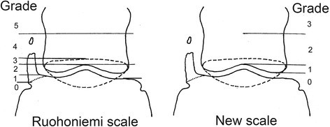
Fig. 2 The OUC grading systems. Grading of ossification according to the Ruohoniemi et al.8 scale (RS) and a new scale (NS) developed in this study. Left side shows moderate ossification with separate center of ossification and the dotted line is the navicular bone. Grading according to Ruohoniemi et al. Grade 0 No ossification, inclination sagittal. Grade 1 Ossification maximum to a level of palmar distal phalanx joint space. Grade 2: To a level of proximal palmar distal phalanx joint space. Grade 3: To a level of proximal border of navicular bone (dotted line). Grade 4: Proximal navicular bone up to distal half of middle phalanx. Grade 5: Ossification over distal half of middle phalanx. New grading according to SLU (new): Grade 0: Ossification not extending proximal of distal middle phalanx. Grade 1: Ossification extending between distal middle phalanx to a level of proximal palmar distal phalanx joint space. Grade 2: To a level of distal half of middle phalanx. Grade 3: To a level over distal half of middle phalanx.
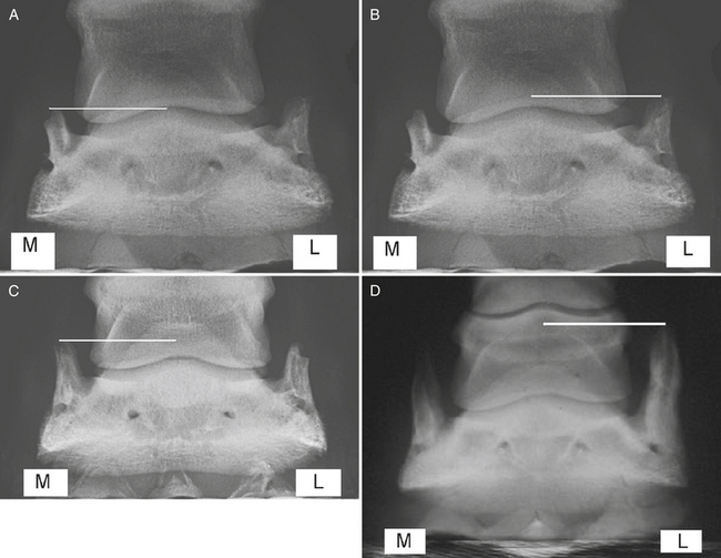
Fig. 3 Dorsopalmar radiographic view of a foot (medial is to the left in A–D) with grade 2 ossification of the medial cartilage of the foot (white line). B, Dorsopalmar radiographic view of a foot with grade 3 ossification of the lateral cartilage of the foot (white line). C, Dorsopalmar radiographic view of a foot with grade 4 ossification of the medial and lateral cartilages of the foot (white line). D, Dorsopalmar radiographic view of a foot with grade 5 ossification of the lateral cartilage of the foot (white line). M, Medial; L, lateral.(Reproduced from Veterinary Key)
Traditional and Specific Shoeing and Farriery for Sidebone
This is often where the art meets the science. Traditional farriery textbooks (e.g. Hickman’s Farriery, Corrective Farriery by Simon Curtis, Principles of Farriery by Colles, Ware & Hayes) contain case reports and detailed guidance for sidebone, often more nuanced than many veterinary articles. Here are refined methods, based on these sources plus practitioner case studies, beyond “bar shoes = answer”:
Key shoemaking / shoeing principles specific to sidebone
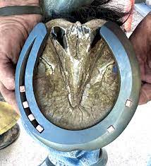
Fig. 4a The traditional Sidebone shoe designed to mimick existing shoe wear with a rolled lateral branch and toe. (Craig Trinka)
- Mimicking existing wear patterns
When a horse has been in shoes, it develops wear that the horse has tolerated. For example, if one side branch (medial or lateral) of the shoe has been worn more, retaining that asymmetry (but refining and balancing it) can reduce sudden stresses. Creating a “sidebone shoe” that is thinner/relieved on the side of ossification (or contralateral to compensate), but supporting the rest of the hoof, helps reduce jarring of the ossified cartilage. - Wide Web / Wide Branch Shoes
Use of wide web keg shoes or hand-forged equivalents that increase the ground-bearing area, especially at the heels, reduces pressure on the collateral cartilages by distributing load more evenly. A wide branch (heel-to-to-toe) helps with heel expansion and reduces lateral bending forces on the ossified cartilage. Videos and demonstrations (e.g. “Sidebone Shoe using Delta Wide Web Keg Shoe”) show these being used successfully for unilateral sidebone. - Relief of affected branch
The shoe may be relieved (thinned) or ground away on the branch over the sidebone to lessen impact/jolt. This means that that branch does less contact with hard ground, reducing direct concussion to the ossified cartilage. - Modified nails / fewer nails on affected side
To avoid pinching or adding rigidity, nails may be placed more upright, or fewer nails to avoid heavy stress or micro-fracture risk in ossified cartilage and its attachments. Sometimes only two nails on the toe branch for the affected side, plus more support medially/laterally via web or pad rather than nails. - Toe roll / Rocker / Breakover modification
Rolling or rocking the toe (shortening the functional lever arm) allows breakover to occur earlier, reducing forward-push forces that can aggravate palmar/heel strain and thus lessen transmitted shock to the collateral cartilages and coffin joint. - Heel height moderation and balancing medial-lateral toe-heel lengths
Restoring or maintaining a correct hoof-pastern axis is vital. Underrun heels or overlong toe increase leverage and increase concussion. Ensuring heels are not too low or excessively trimmed, but neither too high (which can introduce other problems) is part of the balance. Also balancing side-to-side: the medial and lateral heels should be level if conformation allows.
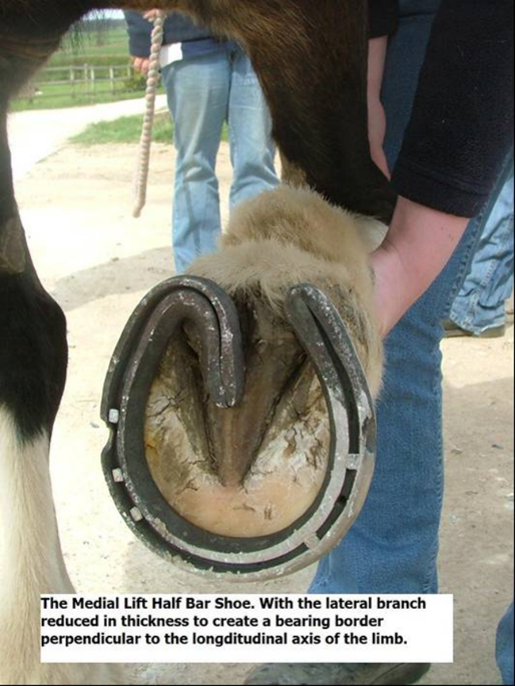
Fig 4b Modifications to the traditional sidebone shoe can include heel height adjusment in the form of a medial spiraling lift. In this case a half frog piece has been added to aid hemodynamic function.
- Pads, packing, and shock-absorbing materials, when needed
In horses with thin soles or where heel conformation is poor, use of pads (leather, cork, synthetic), or packing under the shoe might help cushion impact. Frog support may also be added. However, the pad should not restrict natural heel expansion unduly: rigid pads that interfere with expansion can do more harm. - Cyclical trimming/shoeing intervals
More frequent cycles may be better (e.g. 4-5 weeks rather than every 6-8 weeks) to prevent large compensatory hoof changes between shod states, especially in high-level horses. - Use of handmade shoes vs keg (machine-made) shoes
Handmade shoes allow more precise shaping to match the horse’s foot—including accommodating peculiar wear, adjusting branch widths, nail-placement, reliefs, etc. Traditional texts describe forging specialized shoes for sidebone: “sidebone shoes.” - Consider bare-foot and transition
In some cases, if hoof wall quality, sole depth and environment allow, transitioning to bare-foot (or minimal-shoe) work on soft terrain can reduce continuous artificial loading. Some owners report improved hoof shape, increased flexibility, and better comfort. But in many high-level horses this is difficult to match required demands.
Examples (Case-derived / Traditional)
A case of a hunter with bilateral sidebone where the farrier fashioned new shoes to mimic the previous worn shoes on each foot (i.e. copying the original branch wear asymmetry), combined with trimming to medial-lateral balance. After a period of adjustment, the horse returned to full work.
- The “sidebone shoe” described by Dan Cook: fullered inside, punched outside, branch relief, specific steel bar stock, to deliberately reduce weight and impact on the affected branch.
- Use of a wide-web keg shoe modified by grinding and customising branch widths, combined with rocker toe, in an animal with unilateral sidebone.
- Traditional shoeing texts (e.g., Hickman’s Farriery) stress that shoe fit must follow hoof shape, accommodating where the horse naturally bears, not forcing an ideal symmetrical template when the conformation already is asymmetrical, particularly in cases where collateral cartilages are ossified and rigidity or distortion exist.
Management, Integration with Veterinary Care, and Return to Performance
Complementing shoeing, management includes:
- Controlled exercise progression: begin on soft ground, gradually increase to harder surfaces so that bone, cartilage, and soft tissues can adapt.
- Anti-inflammatory therapy during flare-ups (systemic or local) when imaging or blocks localise pain.
- Rest or reduction in workload, especially during fracture or acute pain episodes.
- Monitoring hoof shape regularly: observe changes in heel expansion, hoof wall flare, medial-lateral balance, sole depth.
- Nutrition to support hoof quality and bone health (minerals, proteins, etc).
- Footing/surface management: softer arenas, avoiding repeated work on hard roads or uneven, concussive surfaces.
For high-level dressage horses, returning to pre-condition performance depends on:
- The degree of damage and involvement beyond sidebone (joints, tendon, cartilage).
- How early and well the intervention was made.
- The horse’s ability to adapt (genetics, hoof quality, conformation).
- Consistency of farrier and veterinary care.
- Time: it may take many months of careful work to restore subtle degrees of flexibility, symmetry, and performance.
Many such horses can return to previous levels, though anecdotally riders/farriers report small-but-real deficits: less suppleness in the heel, slightly reduced shock absorption, more fatigue on hard surfaces, occasional sensitivity in collection or lateral work. These may not be visible in low-level performance but matter in high dressage.
Why This Pathology is Overlooked & Why It Matters
Sidebone is easy to ignore because:
- Ossification may be silent for years.
- Radiographs are routinely taken in horses for other reasons and sidebone shows up as an incidental finding.
- Veterinary literature often treats it as “rarely causing clinical lameness,” which is partly true—but that underplays subtle performance impairment.
But for the scientific and performance-horse community this matters because movement symmetry, hoof expansion, shock absorption, caudal limb health and fine joint function (coffin joint, navicular), all depend on what happens at the heel—where sidebone sits. Dressage demands precision, suppleness, quietness of transitions, ability to be light behind. If the heel is stiff or pain-sensitive, that undermines many foundational movements.
Encouragement for Further Research & Study
Traditional farriery sources remain rich with experiential detail, but rigorous controlled studies on sidebone are sparse. Questions ripe for investigation:
- How much residual performance loss is quantifiable (stride length, symmetry, kinetics) in horses with stable ossification versus those without.
- Comparative trials of different shoeing modalities for sidebone (custom sidebone shoes, wide web modifications, relief of one branch, etc).
- Imaging biomarkers (MRI, CT) that predict poorer prognosis.
- Long-term hoof shape and mechanical effects (e.g. changes in heel width, collateral cartilage stress) under different trimming/shoeing regimens.
Conclusion
Sidebone is more than a “radiograph curiosity.” For many horses it remains silent—but for others, especially high-performance or conformed horses, it can lead to subtle but meaningful performance compromises or overt lameness. Traditional farriery offers a variety of refined shoeing strategies—mimicking wear, relieving affected branches, using wide webs, adjusting breakover—that can make a real difference. With early detection, thoughtful veterinary diagnosis, skilled farriery, consistent hoof care and realistic expectations, many horses can and do return to high levels of work. And even where perfect return isn’t possible, improvement in comfort, symmetry, and longevity of soundness is often achievable.
If you’re intrigued, I suggest studying Corrective Farriery (Vol I & II, Simon Curtis), Hickman’s Farriery, and Principles of Farriery (Colles, Ware & Hayes), seeking out case studies of “sidebone shoes,” and observing in practice how nuanced modifications of branch width, relief, fit, and nail placement can shift outcomes.
Selected References
- Curtis, S.J. (2002/2006) Corrective Farriery: A Textbook of Remedial Horseshoeing Volumes I & II.
- Hickman, J. (revised edition) Hickman’s Farriery: The Complete Guide to Horseshoeing.
- Colles, C., Ware, R. & Hayes, J. (2022) Principles of Farriery.
- O’Grady, S.E. (2008) “Basic Farriery for the Performance Horse”, Veterinary Clinics: Equine Practice, 24(1), pp. 203-218.
- “Making the Sidebone Shoe” (Cook, D.), American Farrier’s Journal, 1999.
- Seery, S. (2021) Hoof shape and loading in sound and lame horses: how this is influenced by farriery. PhD thesis, University of Liverpool.