
Proportional Radiographic Relationships Within the Equine Foot
Proportional Radiographic Relationships Within the Equine Foot
Andrew Prout AWCF. Feet First Farriery Limited. Welltown Cottage, Welltown, Cardinham, Bodmin, Cornwall. PL30 4EG
Abstract
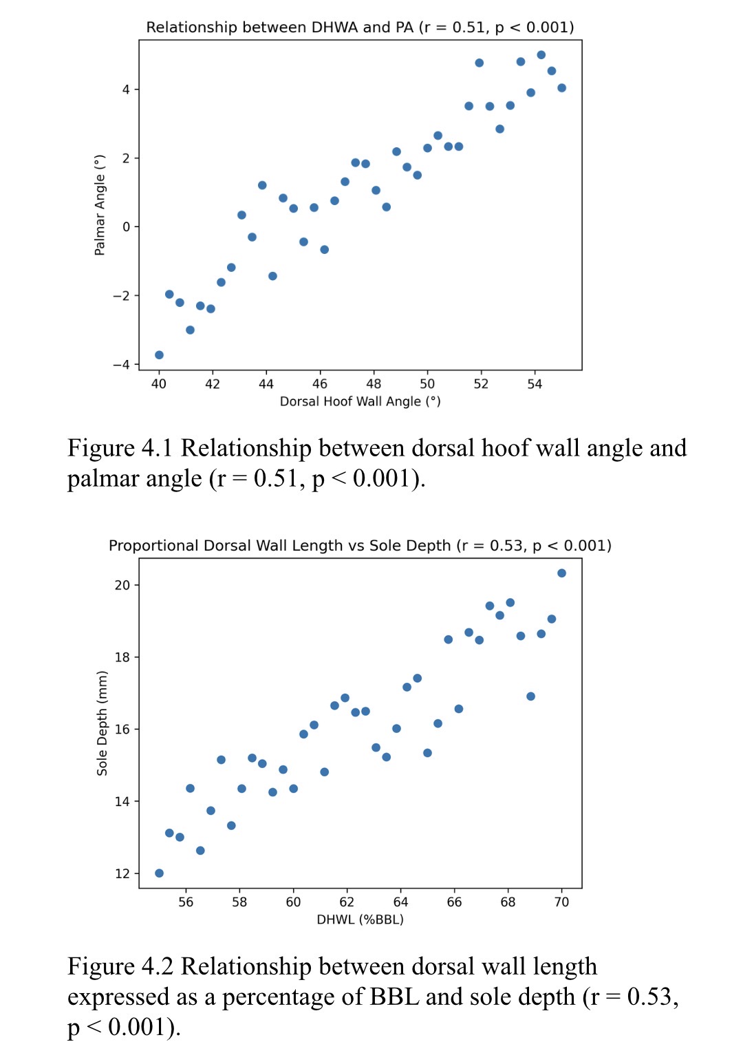
This research investigates proportional radiographic relationships within the equine foot using blinded, randomly supplied lateromedial radiographs. Linear measurements were normalised to bearing border length (BBL) of the distal phalanx to control for inter-individual variation in foot size. Relationships between dorsal hoof wall angle (DHWA), palmar angle (PA), sole depth (SD), and proportional internal anatomy were examined using descriptive statistics, Pearson correlation analysis with false discovery rate correction, principal component analysis (PCA), and multivariable regression. Significant associations were identified between DHWA and PA, and between proportional dorsal hoof wall length and sole depth. Hoof lamellar zone (HLZ) measures were highly intercorrelated, indicating a coherent dorsal wall–P3 interface. These findings demonstrate that the equine foot functions as an integrated proportional system with direct relevance to evidence-based farriery practice.
Screenshot
References
Bowker, R.M. (2003) The growth and adaptive capabilities of the hoof wall and sole. Equine Veterinary Journal, 35(3), pp. 258–264.
Coleman, E. (1802) Observations on the Structure and Diseases of the Foot of the Horse. London: T. Cadell.
Dollar, J.A.W. (1898) A Handbook of Horseshoeing. London: Baillière, Tindall and Cox.
Dyson, S.J. and Murray, R. (2007) Management of foot balance. Equine Veterinary Education, 19(10), pp. 531–540.
Parks, A.H. (2011) Form and function of the equine digit. Veterinary Clinics of North America: Equine Practice, 27(1), pp. 1–17.
Russell, W. (1879) Scientific Horseshoeing. New York: R.M. McBride.