
Navicular Syndrome in the Horse: Aetiology, Diagnostics, and Multimodal Treatment Strategies Including Farriery Approaches
Caldwell M.N. & Nadden N. HoofFlix. Com., 116, Newcastle Road, Talke, Staffordshire. ST71SA email info@hoofflix.com
Abstract
Navicular syndrome represents a multifactorial pathology of the equine foot, most commonly affecting sport and leisure horses. Once considered a degenerative condition isolated to the navicular bone, it is now recognised as a disease complex involving soft tissue structures of the podotrochlear apparatus, including the deep digital flexor tendon (DDFT), navicular bursa, and supporting ligaments. Advances in imaging—especially magnetic resonance imaging (MRI)—have revolutionised diagnosis, shifting treatment strategies toward more targeted and biomechanically rational interventions. This paper explores current theories of aetiology, diagnostic protocols, and treatment options, with a specific emphasis on the mechanical rationale for contemporary farriery treatments and the influence of hoof conformation on pathology, referencing the pivotal findings of Caldwell (2017).
Introduction
Navicular syndrome is one of the leading causes of chronic forelimb lameness in horses, particularly among performance breeds (Dyson et al., 2018). Traditionally viewed as an osteoarthritic condition of the navicular bone, it is now widely understood to involve a spectrum of pathologies encompassing both osseous and soft tissue components of the podotrochlear apparatus (Schramme et al., 2020). This evolving understanding has been driven by advanced imaging, primarily MRI, which permits detailed evaluation of internal foot structures in vivo (Denoix, 2019). Recognition of the biomechanical stresses affecting the navicular region has also shifted the therapeutic emphasis toward corrective farriery that modifies loading patterns within the foot.
Relevant Anatomy: The Podotrochlear Apparatus
The navicular apparatus consists of the navicular bone (distal sesamoid), navicular bursa, deep digital flexor tendon (DDFT), collateral sesamoidean ligaments, impar ligament, and associated soft tissues (Eliashar, 2007). These structures function together to absorb and distribute biomechanical load through the palmar aspect of the distal limb during weight-bearing and breakover. Disruption to any of these components may contribute to the cascade of navicular pathology.
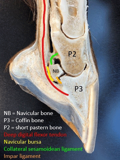
Illustration 1. Sagittal MRI section of the equine foot showing the navicular bone, DDFT, and navicular bursa.
Aetiology and Risk Factors
Navicular syndrome is now considered a consequence of chronic mechanical overloading, leading to both soft tissue strain and bone remodelling. Several predisposing factors have been identified, including poor hoof conformation, long toe–low heel structures, broken-back hoof-pastern axis, and small or contracted feet (Caldwell, 2017; Dyson et al., 2011). Horses working on hard or repetitive surfaces are also at increased risk due to higher concussive loading on the palmar foot.
Hoof morphology plays a pivotal role. Caldwell’s (2017) doctoral thesis established strong correlations between hoof shape—especially dorsopalmar imbalance—and pathological MRI findings, including DDFT lesions and navicular bone sclerosis. This evidence supports a biomechanical model of disease in which hoof proportions alter the direction and magnitude of forces acting on the podotrochlear structures, particularly the navicular bone and adjacent soft tissues.
Clinical Signs and Diagnostic Workup
Affected horses often present with intermittent or progressive unilateral or bilateral forelimb lameness that worsens on hard ground or when circling. Hoof testers may elicit pain over the frog or heels, and gait evaluation often reveals shortened cranial phase or toe-first landing.
Localisation through diagnostic anaesthesia typically begins with palmar digital nerve blocks. A positive response may indicate navicular involvement, but this block also desensitises adjacent structures, necessitating further investigation (Schramme et al., 2020).
Radiographs remain a first-line imaging tool, although early or soft tissue-dominant cases may yield minimal findings. Common radiographic indicators include flexor cortex lysis, synovial invaginations, or medullary sclerosis (Denoix, 2019). However, these changes often appear late and do not reliably correlate with clinical severity.
Magnetic resonance imaging (MRI) is now the gold standard for navicular diagnosis. It allows direct evaluation of both bone and soft tissue, including DDFT fibre disruption, bursal distension, adhesions, and navicular bone oedema (Murray et al., 2006). These findings are critical for establishing the primary source of pain and selecting appropriate treatment.
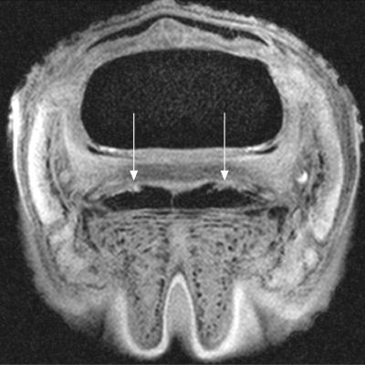
Illustration 2. MRI case showing DDFT dorsal fibre tearing adjacent to the navicular bursa.A typical presentation of tendon-associated navicular syndrome.
Veterinary Treatment Strategies
Medical management is generally aimed at reducing inflammation and improving circulation within the navicular region. Non-steroidal anti-inflammatory drugs (NSAIDs) are widely used for pain control but do not alter disease progression. Intra-bursal corticosteroid injections can provide short-term relief in cases with synovial involvement (Eliashar, 2007).
Bisphosphonates such as tiludronate or clodronate have shown promise in reducing bone resorption and pain associated with navicular bone changes (Haussler et al., 2021). In selected cases, regenerative therapies—such as platelet-rich plasma (PRP) or stem cells—have been explored, particularly in horses with tendon-related pathology identified on MRI.
Farriery Treatment: Mechanical Rationale and Clinical Application
Farriery forms the cornerstone of long-term management for navicular syndrome and must be customised to the individual’s hoof morphology and clinical findings. The primary biomechanical goals are to:
Reduce strain on the DDFT and navicular apparatus during breakover,
Restore hoof-pastern axis alignment,
Improve load distribution across the palmar foot,
Enhance shock absorption and digital cushion function.
Trimming is the first line of correction. Caldwell (2017) emphasises restoring the appropriate dorsal hoof wall angle, reducing toe length, and gradually raising underrun heels to shift loading palmarly. This improves the orientation of the DDFT over the navicular region, reducing compressive and shearing forces.
Shoe selection is critical. Rolled or rockered toe shoes ease breakover, while heeled shoes (e.g., wedge pads or raised heels) temporarily reduce tension on the DDFT, particularly during rehabilitation. Wide-webbed or bar shoes increase the ground contact area and frog engagement, assisting in distributing load away from the damaged region (O’Grady and Poupard, 2003). Some cases benefit from shock-absorbing materials, such as pour-in pads or composite shoes.
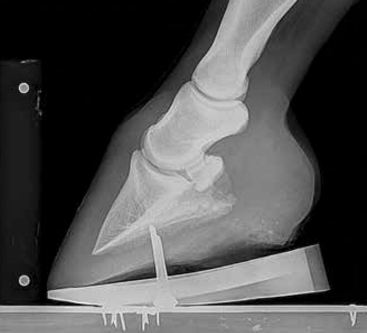
Illustration 3. shieing strategies are based in phalngeal alignment, extension of the palmar bearing surface of the hoof and restoration of HPA, relief of tension of the DDFT at the site of the DS and protection from external violence over the anatomical region of the DS. Each of these interventions should match individual clinical signs.
findings. For example, in horses with confirmed DDFT tears, raising the heel temporarily reduces DDFT tension during healing. Conversely, horses with isolated bone changes but no soft tissue damage may benefit more from lateral ground surface expansion or bar shoes with pads.
Prognosis and Rehabilitation
Prognosis varies depending on the structures involved, the chronicity of the condition, and the success of farriery intervention. Horses with primarily bony changes and minimal soft tissue disruption may return to full work, particularly when conformation is corrected early. However, those with advanced DDFT lesions or chronic navicular bursitis often require long-term maintenance therapy and may not return to prior performance levels.
Rehabilitation protocols typically include rest, corrective shoeing, and gradual reintroduction to controlled exercise over several months. Close collaboration between veterinarian and farrier is essential throughout the recovery process.
Conclusion
Navicular syndrome is a complex and multifactorial disease involving both bony and soft tissue structures of the podotrochlear apparatus. Diagnosis has evolved significantly with the advent of MRI, facilitating more specific identification of underlying pathologies. Treatment is most successful when combining medical management with mechanically rational farriery based on individual conformation and biomechanical function. The work of Caldwell (2017) and others has provided robust evidence linking hoof morphology with pathology and underscoring the importance of hoof balance and function in both prevention and recovery.
References
Caldwell, M. N. (2017). The Relationship Between Hoof Morphology and MRI Findings in Equine Foot Pathologies. Doctoral Thesis, University of Liverpool.
Denoix, J. M. (2019). Diagnosis and Management of Lameness in the Horse (2nd ed.). Elsevier.
Dyson, S., Murray, R., Schramme, M., & Blunden, T. (2011). Current concepts of navicular disease. Equine Veterinary Education, 23(1), 27–39.
Dyson, S. et al. (2018). Lameness associated with foot pain: Diagnosis and MRI findings. Equine Veterinary Journal, 50(3), 412–426.
Eliashar, E. (2007). The biomechanics of the equine digit and its relevance to the diagnosis and treatment of lameness. Equine Veterinary Journal, 39(1), 61–66.
Haussler, K. K., et al. (2021). Clinical efficacy of bisphosphonates in managing navicular disease. Journal of Equine Veterinary Science, 96, 103284.
Murray, R. C., Dyson, S. J., Tranquille, C. A., & Adams, V. (2006). Use of MRI for the investigation of foot lameness. Equine Veterinary Education, 18(6), 335–342.
O’Grady, S. E., & Poupard, D. A. (2003). Foot care for horses with heel pain. Veterinary Clinics of North America: Equine Practice, 19(2), 365–385.
Schramme, M. C., et al. (2020). Advances in diagnostic imaging of the equine foot. Veterinary Radiology & Ultrasound, 61(4), 393–405.