
Farriery Radiographs & Foot Mapping
Radiographs and Foot Mapping Keep Your Horse’s Hooves Healthy
Introduction
Maintaining hoof balance is essential for equine soundness. Even superficial hoof issues can mask deeper imbalances within the hoof capsule. Radiographs—commonly X‑rays—provide a window into the bones and soft tissues beneath, enabling veterinarians and farriers to plan trims and shoes with precision. Regular foot mapping, as developed by Dr Mark Caldwell, further refines this process: by documenting external hoof geometry, mapping supports early detection of imbalance and drives timely radiographs. This approach helps negate the cost associated with reactive imaging and ensures interventions are targeted.
Why Foot Balance Matters
A well-balanced hoof distributes weight evenly, minimizing stress on joints, tendons, and soft tissue structures (Dyson & Murray, 2007)
Misalignment—though occasionally imperceptible from the outside—can lead to lameness, abnormal wear, and chronic pathology. Foot mapping provides consistent external reference points, helping practitioners detect asymmetries before internal damage occurs.
What Is Radiography of the Hoof?
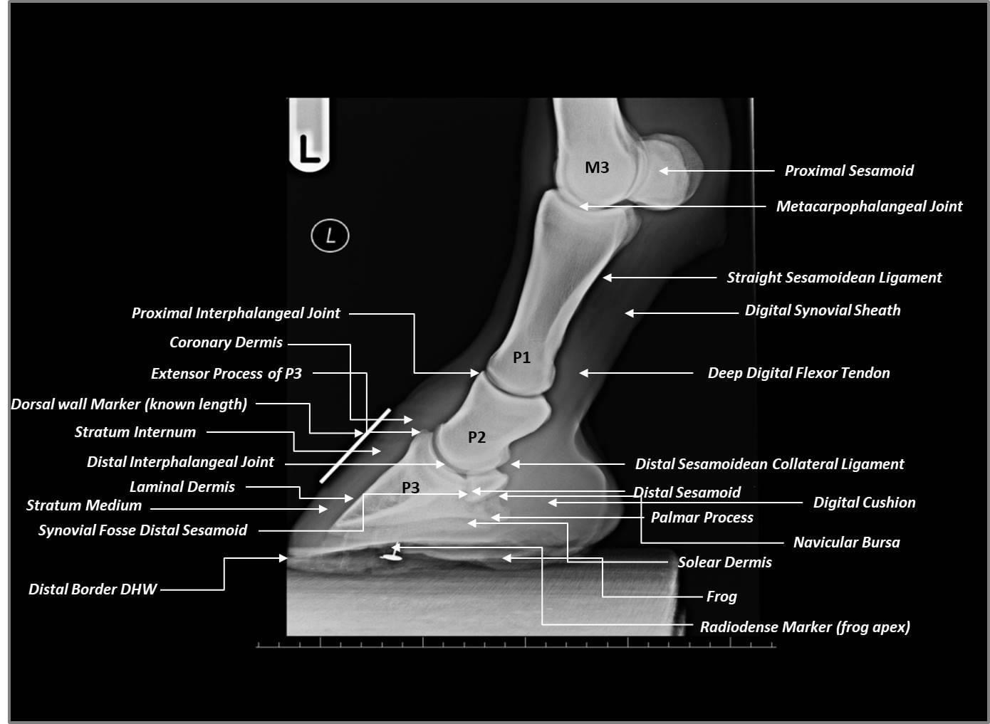
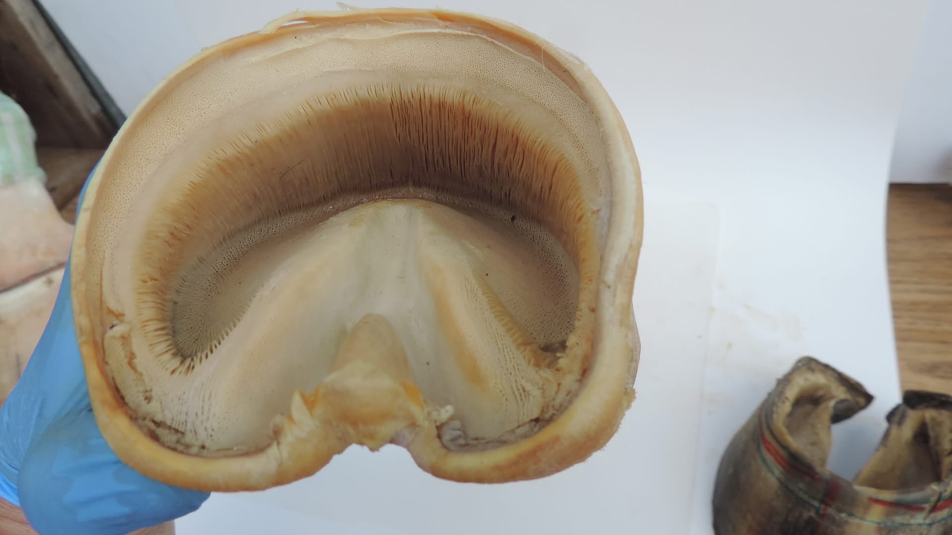
Radiographs use low-dose radiation to visualize internal anatomy—specifically the coffin bone (P3), navicular bone, and pastern bones—while the horse stands (White, 2024)The Horse. Most equine radiographs require no sedation and are highly informative when properly executed.
Key Radiographic Views:
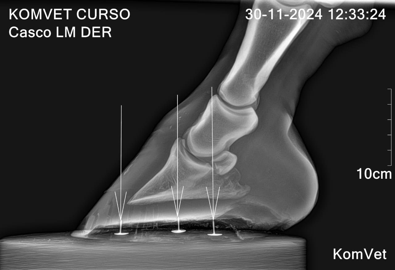
Lateromedial (LM): side view showing toe length, heel height, and hoof-pastern axis.
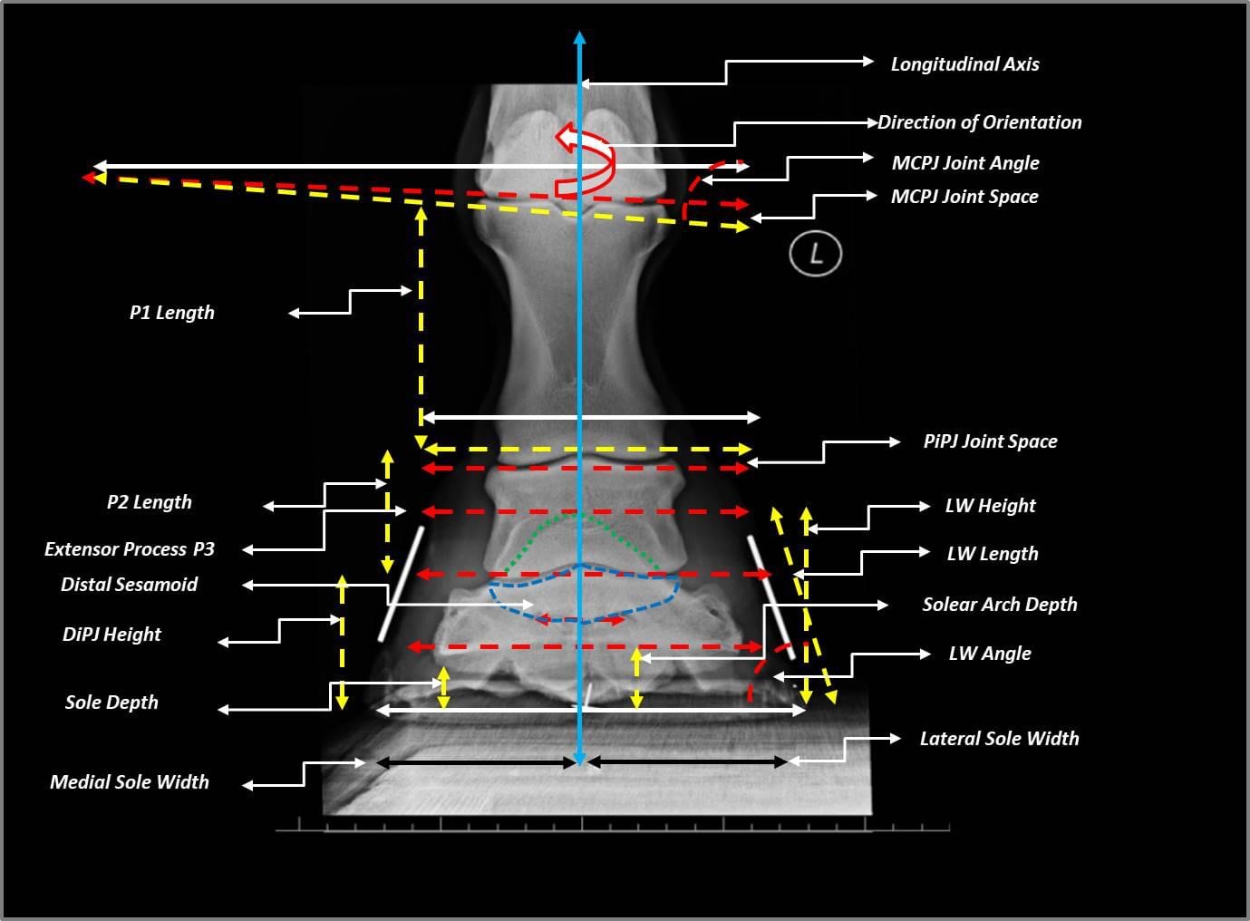
Dorsopalmar (DP): front view assessing side-to-side balance.
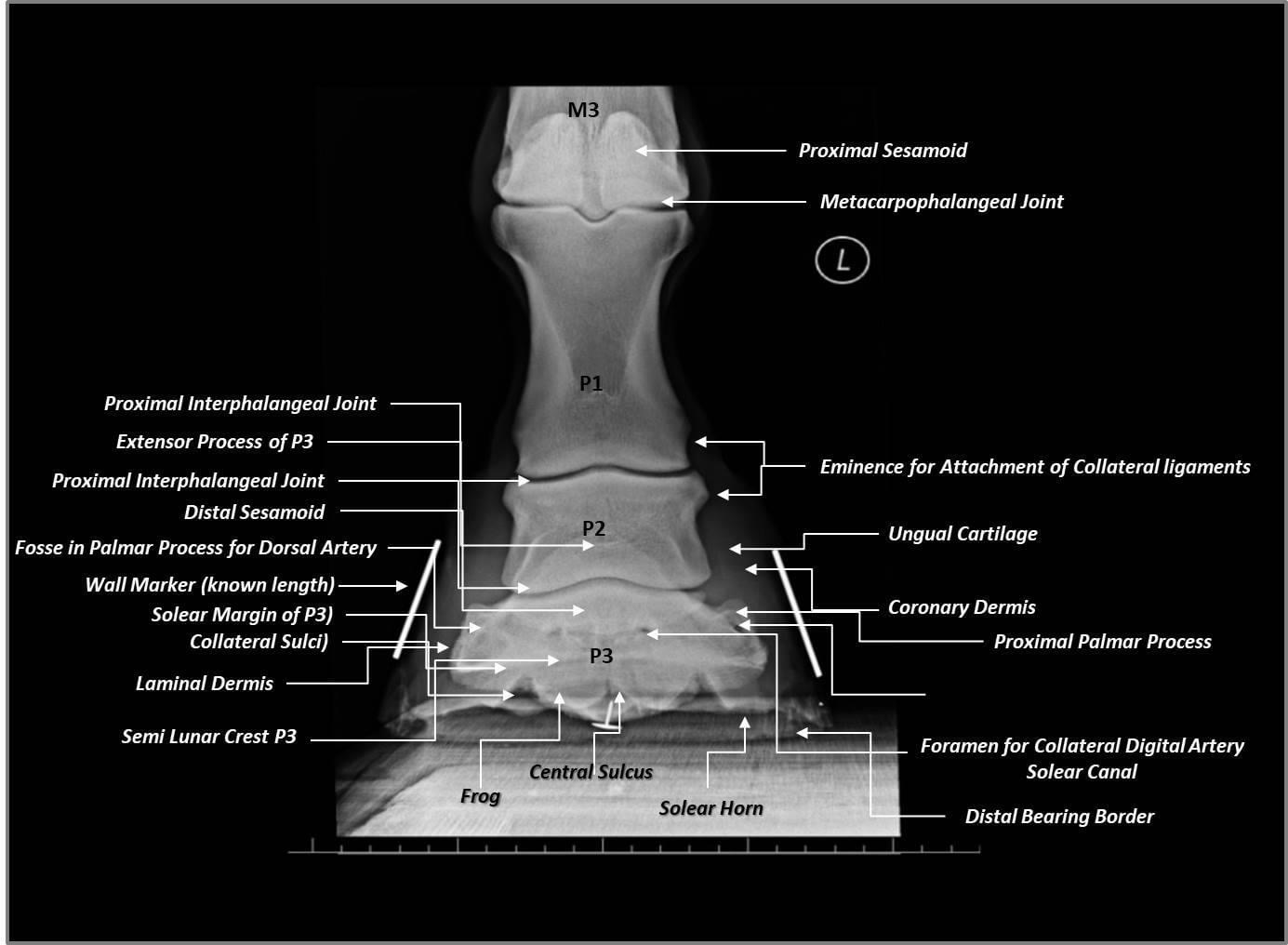
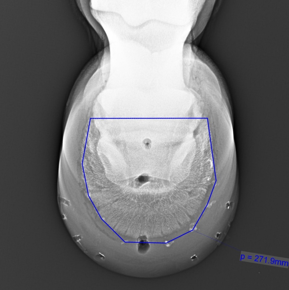
Solar (broad sole view): checks sole thickness and pathology like abscesses.
Each view contributes crucial data for trimming and shoeing decisions.
Radiographic Indicators of Hoof Balance
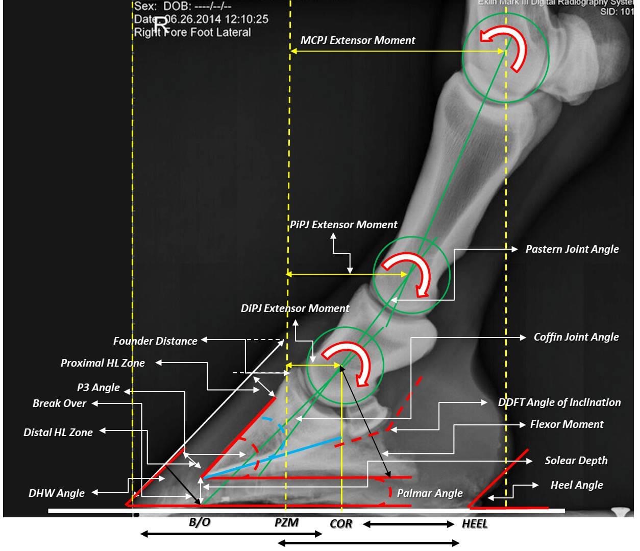
1. Hoof‑Pastern Axis (HPA)
An aligned HPA means the hoof wall matches the pastern angle. A “broken-back” axis signals excessive toe length or under-run heels, increasing stress on the deep digital flexor tendon and navicular region (Clayton et al.,2011)
2. Palmar Angle (PA)
This angle, between the ground and the coffin bone’s solar surface, should typically measure +2° to +5° in a healthy front foot. Negative or low angles indicate heel collapse and increased risk of lamellar disease (Kummer et al., 2006)americanfarriers.com.
3. Mediolateral Balance
From the DP view, veterinarians can detect asymmetry that may strain internal structures. Significant side-to-side discrepancies are often associated with limb conformation or uneven wear (Weller et al., 2006)
4. Sole Depth
Sole measurements under 10–12 mm increase the risk of bruising and pain. Radiographs help determine when reinforcement or protective trimming is needed (Dyson, 2011)americanfarriers.com.
How Regular Foot Mapping Enhances Radiograph Use
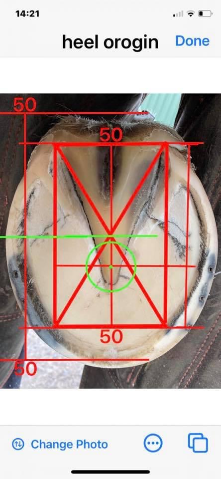
Dr Mark Caldwell’s foot mapping protocol uses external landmarks to pinpoint internal structures such as the centre of rotation (CoR) and break‑over point (Bo) using grid lines projected from heel buttresses over the frog apex (Caldwell et al. 2015)
This enables farriers to plan trimming and shoe placement precisely—even before imaging.
Benefits:
Reduces unnecessary radiographs by tracking external hoof changes.
Enables early detection of imbalance, prompting timely imaging.
Offers a cost‑effective way to monitor hoof geometry between radiographs.
Supports accurate radiographic interpretation by correlating external and internal coordinates (Moon 2017
Integrating Radiographs & Foot Mapping to Guide Hoof Care
Step‑by‑Step Protocol:
Map the hoof: mark CoR, CoP, frog apex, and break‑over externally.
Observe changes over time: record hoof geometry monthly.
Trigger radiographs when imbalances appear on mapping grid or the foot: either at baseline, with clinical signs, or alongside seasonal farriery cycles.
Interpret LM and DP radiographs using fixed scale markers for accurate measurements (Eggleston, 2012)
Plan trimming/shoeing that restores proper HPA, palmar angle, and CoR alignment.
Practical Examples of Hoof Radiograph Guidance
Rotated coffin bone—trimming shortens toe to reduce leverage.
Collapsed heel/palmar angle—shoeing or wedge support may raise heel to restore angle.
Uneven mediolateral distribution—side-specific relief can be provided.
Thin sole—minimal trimming and protection pads are selected.
Even in sound horses, baseline radiographs help tailor long-term hoof care and prevent costly structural issues (White, 2024)
Limitations and Best Practices
Radiographs are two-dimensional snapshots of a three-dimensional structure. Misalignment or poor positioning can skew results (Butler et al., 2008)NANRIC. Proper technique—including foot placement, scale markers, and dorsal wall alignment—is essential (Equipodiatry 2021)Mapping alone is not definitive; it complements radiographic assessments, not replaces them.
Conclusion
Radiographs, when used thoughtfully, are powerful tools in hoof care—especially when timed and interpreted alongside regular foot mapping. Dr Caldwell’s mapping method provides an affordable means to track hoof balance, triggering interventions early and reducing radiograph dependency. Through this integrated system, veterinary and farrier teams can work collaboratively to ensure hoof health, prevent disease progression, and maintain long-term soundness.
References
Butler, J.A. et al. (2008) Clinical Radiology of the Horse, 3rd ed., Wiley‑Blackwell.
Clayton, H.M. et al. (2011) ‘Kinematics and kinetics of the hoof and the hoof‑pastern axis’, Equine Veterinary Journal, 43(3), pp. 278–285. americanfarriers.com
Caldwell, M.N. et al. (2015) ‘Validating external hoof mapping against internal geometry’, Forge Knowledge, 33(11), pp. 34–36.
Dyson, S. (2011) ‘Diagnosis and management of common foot problems in the horse’, Equine Veterinary Education, 23(4), pp. 178–189. americanfarriers.com
Dyson, S.J. & Murray, R.C. (2007) ‘Management of equine foot pain’, Vet Clin North Am Equine Practice, 23(2), pp. 417–446. americanfarriers.com
Eggleston, R.B. (2012) ‘The Value of Quality Foot Radiographs’, Equipodiatry.com. equipodiatry.com+1The Horse+1
Kummer, M. et al. (2006) ‘Effect of hoof trimming on radiographic parameters’, Veterinary Journal, 172(1), pp. 58–66.ResearchGate
Moon, G. (2017) Hoof mapping accuracy evaluation, Fellowship manuscript, British Farrier Journal. wcf.org.uk
White, C. (2024) ‘The Value of Hoof Radiographs’, TheHorse.com, Oct 15. The Horse
Weller, R. et al. (2006) ‘Variation in conformation’ Equine Veterinary Journal, 38(S36), pp. 616–621.americanfarriers.com
By combining foot mapping with strategic radiographic imaging, professionals can adopt a highly informed, cost-effective hoof-care program that optimises alignment, function, and long-term horse wellbeing.