
Current Scientific Understanding of Equine Hoof Expansion, Stance Phase Loading, and Conformational Influences
Caldwell M.N., Madden N.
HoofFlix.com (info@hoofflix.com)
key words;
The traditional theory of hoof expansion posits that during weight bearing—particularly mid-stance to terminal stance—the caudal hoof structures, including the quarters and heels, undergo lateral splaying or “expansion” to accommodate ground reaction forces (GRFs), dissipate shock, and facilitate venous return through compression of the digital cushion and frog (Bowker et al., 1998; O’Grady & Poupard, 2003). However, recent biomechanical studies suggest this expansion is both less dramatic than once thought and highly dependent on dynamic load vectors, conformation, and surface interaction.
1. Hoof Expansion Mechanics and Stance Phase Loading
The stance phase of the equine stride is divided into four biomechanical stages: initial contact, loading response, midstance, and terminal stance. The point and angle of force application throughout these phases significantly influences hoof deformation. When the hoof lands flat or slightly heel-first, the posterior structures (heels, frog, bars)are preferentially loaded, which may facilitate moderate lateral deformation or “expansion” (Clayton et al., 2011).
Conversely, toe-first or mediolateral imbalanced landings alter the location of peak vertical force, leading to asymmetric hoof distortion (Hagen et al., 2020). Excessive force directed toward one side of the hoof increases strain on laminar attachments and can cause non-physiological capsule deformation over time, including sheared heels and displaced bars (Eliashar et al., 2004).
Key Point:
Hoof expansion is not a uniform outward movement but rather a complex, strain-distribution response to cyclical loading forces. Its magnitude and symmetry depend heavily on the landing pattern and force vector orientation during stance (Van Heel et al., 2005).
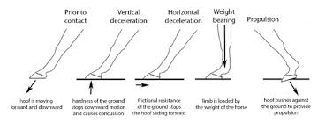
Figure 1: GRF vectors and hoof contact zones at
2. Influence of Digital and Hoof Capsule Conformation
Conformation plays a critical role in how load is transferred through the hoof and whether expansion occurs symmetrically or pathologically. Horses with upright pastern angles, club feet, or negative plantar/dorsal angles demonstrate altered force dissipation through the hoof capsule, resulting in reduced expansion and/or asymmetrical distortion (Dyson et al., 2011).
Club foot conformation results in a dorsally displaced centre of rotation and increased load on the dorsal lamellae, with reduced engagement of the frog and digital cushion—thus limiting caudal expansion (Dik & van den Broek, 1995).
Underrun heels or negative palmar/plantar angles shift the load anteriorly during midstance, creating excessive stress at the toe and proximal interphalangeal joint, while simultaneously leading to collapsed caudal structureswith little or no elastic deformation (O’Grady & Parks, 2008).
In horses with long-toe, low-heel conformations, the mechanical axis of loading is shifted forward, often reducing dynamic expansion and increasing shear stress at the distal interphalangeal joint and navicular region (Kane et al., 1998).
Furthermore, the horn tubule orientation and sole concavity influence how strain is distributed under compressive loads. In healthy feet, the tubules bend slightly to absorb force and rebound elastically. In diseased or poorly conformed feet, this property is compromised, reducing both expansion and shock absorption (Pollitt, 1990).
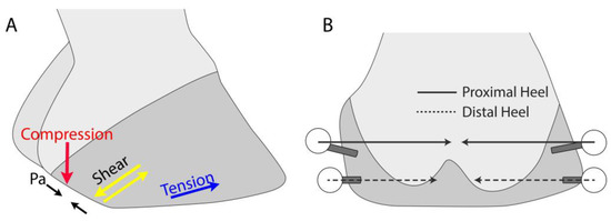
Figure 2: Comparative diagrams of hoof s principle strains and direction that ultimately lead to underrun conformations.
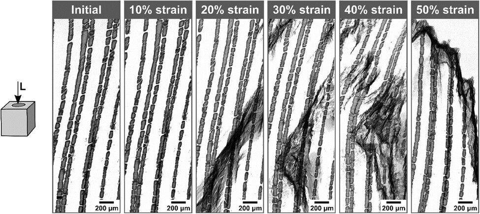
Figure 3:shows the progression in increments of 10% strain of a dry hoof sample cracking under compressive load. Formation of large black splotching corresponds to cracks weaving between the tubules and tubule bridges, initiating between 10% and 20% strain. Tubules begin to bend toward the top of the field of view at around 30% strain until a clean fracture occurs at 50% strain, shearing off the tops of the tubular cavities. (Lazarus et al 2023).
3. Venous Return and Expansion: Reassessing the Hydraulic Theory
The hydraulic theory of hoof expansion asserts that the cyclic compression of the frog and digital cushion during stance facilitates venous return from the foot (Hood, 1982). While widely cited, this concept has been challenged in recent years due to lack of direct hemodynamic evidence. A study by Licka and Peham (2011) showed limited evidence of significant internal volume change in the hoof capsule during the stance phase, suggesting that hoof expansion may be less critical to circulation than once believed, particularly in shod horses.
However, proper caudal engagement does appear to influence tissue perfusion and metabolic waste removal through intermittent pressure fluctuations, supporting the rationale for ground-contacting frog and sole engagement in both barefoot and correctly shod feet (Bowker, 2003).
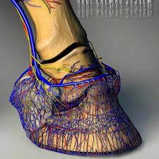
Figure 4. Complex and intricate vascular supply of the equine hoof
4. Implications for Farriery
From a farriery perspective, promoting balanced, symmetrical loading through appropriate trimming and shoeing strategies is essential to maintain healthy hoof expansion dynamics. Key strategies include:
Ensuring mediolateral symmetry in hoof capsule geometry (e.g., quarter wall height, medial/lateral ground surface).
Maintaining proper dorsopalmar balance (based on centre of rotation and palmar angle) to allow normal strain distribution.
Selecting shoeing methods that do not restrict caudal expansion, such as open-heeled or frog-supporting shoes, especially in cases of underrun heels or caudal collapse (Clayton et al., 2011).
Using hoof mapping to guide objective decisions about distortion correction and balance (Caldwell, 2017).
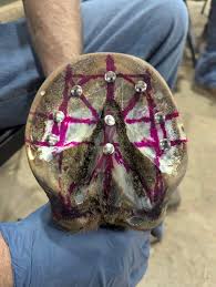
Figure 4: Hoof mapping image showing static imbalance with annotations.
Conclusion
The hoof expansion theory must now be understood not as a simple mechanical splaying, but as a dynamic interaction between conformation, loading vector, and structural hoof integrity. Recognising the biomechanical and anatomical factors that govern expansion during stance is essential for evidence-based hoof care. Farriery strategies should aim to optimise force distribution, prevent asymmetric strain, and maintain healthy digital biomechanics.
References
Bowker, R.M., Van Wulfen, K.K., Springer, S.E., & Linder, K.E., 1998. Functional anatomy of the vascular elastic tissues in the equine foot. American Journal of Veterinary Research, 59(5), pp. 513–520.
Clayton, H.M., Gray, S., Kaiser, L.J., & Bowker, R.M., 2011. Effects of barefoot trimming on hoof expansion. Equine Veterinary Education, 23(11), pp. 527–531.
Dik, K.J. & van den Broek, J., 1995. Club foot in foals: radiographic and clinical characteristics. Equine Veterinary Journal, 27(2), pp. 111–115.
Dyson, S.J., Tranquille, C.A., Collins, S.N., Parkin, T.D.H., & Murray, R.C., 2011. The influence of foot balance on lameness and pathological change. Equine Veterinary Journal, 43(5), pp. 535–543.
Eliashar, E., McGuigan, M.P., Rogers, K.A., & Wilson, A.M., 2004. A finite element analysis of a three-dimensional equine hoof. Equine Veterinary Journal, 36(8), pp. 679–685.
Hagen, J., Baldrighi, J., & Clayton, H.M., 2020. Effect of hoof imbalance on kinetic parameters. Journal of Equine Veterinary Science, 89, 103010.
Hood, D.M., 1982. The mechanism of hoof expansion. Journal of Equine Veterinary Science, 2(3), pp. 123–128.
Kane, A.J., Stover, S.M., Gardner, I.A., Bock, K.B., Case, J.T., Johnson, B.J., Anderson, M.L., Barr, B.C., Daft, B.M., Kinde, H., & others, 1998. Hoof size, shape, and balance as possible risk factors for musculoskeletal injury of Thoroughbred racehorses. American Journal of Veterinary Research, 59(12), pp. 1545–1552.
Licka, T. & Peham, C., 2011. The in vivo volume changes of the equine hoof capsule during loading. The Veterinary Journal, 190(1), pp. 56–61.
O’Grady, S.E. & Parks, A.H., 2008. Farriery for the broken-back hoof-pastern axis. Equine Veterinary Education, 20(1), pp. 24–30.
O’Grady, S.E. & Poupard, D.A., 2003. Footcare for horses with heel pain. Veterinary Clinics of North America: Equine Practice, 19(2), pp. 365–379.
Pollitt, C.C., 1990. The anatomy and physiology of the inner hoof wall. Equine Veterinary Education, 2(2), pp. 48–54.
Van Heel, M.C., Kroekenstoel, A.M., van Dierendonck, M.C., & Barneveld, A., 2005. Uneven feet in a foal. Equine Veterinary Education, 17(4), pp. 199–204.