
Biomechanical and Clinical Rationale for a Modular Therapeutic Clog System in the Management of Equine Laminitis
Biomechanical and Clinical Rationale for a Modular Therapeutic Clog System in the Management of Equine Laminitis
Caldwell M, Madden N and Charnley N. 116, Newcastle Road, Talke, Staffordshire. ST71SA. email info@hoofflix.com
Abstract
Equine laminitis is a debilitating condition resulting from failure of the laminar suspension apparatus of the distal phalanx (P3). This paper presents a comprehensive review of the biomechanical rationale for therapeutic intervention, focusing on a modular two-part clog system. This system is designed to redistribute ground reaction forces, optimise load distribution, and improve hoof stability in laminitic horses. Emphasis is placed on the integration of trimming protocols, deformable EVA footbed material, non-invasive glue application, and the use of removable treatment plates. Biomechanical models, case studies, and clinical outcomes are discussed.
Introduction
Laminitis involves the structural failure of the lamellar interface between the hoof capsule and P3. The displacement of P3 leads to altered biomechanical loading patterns, creating further lamellar failure and associated pain. Conventional approaches focus on unloading the dorsal laminae and controlling displacement. Recent advances in farriery have seen the emergence of modular therapeutic clog systems capable of dynamically adjusting biomechanical forces while facilitating ease of application and monitoring.
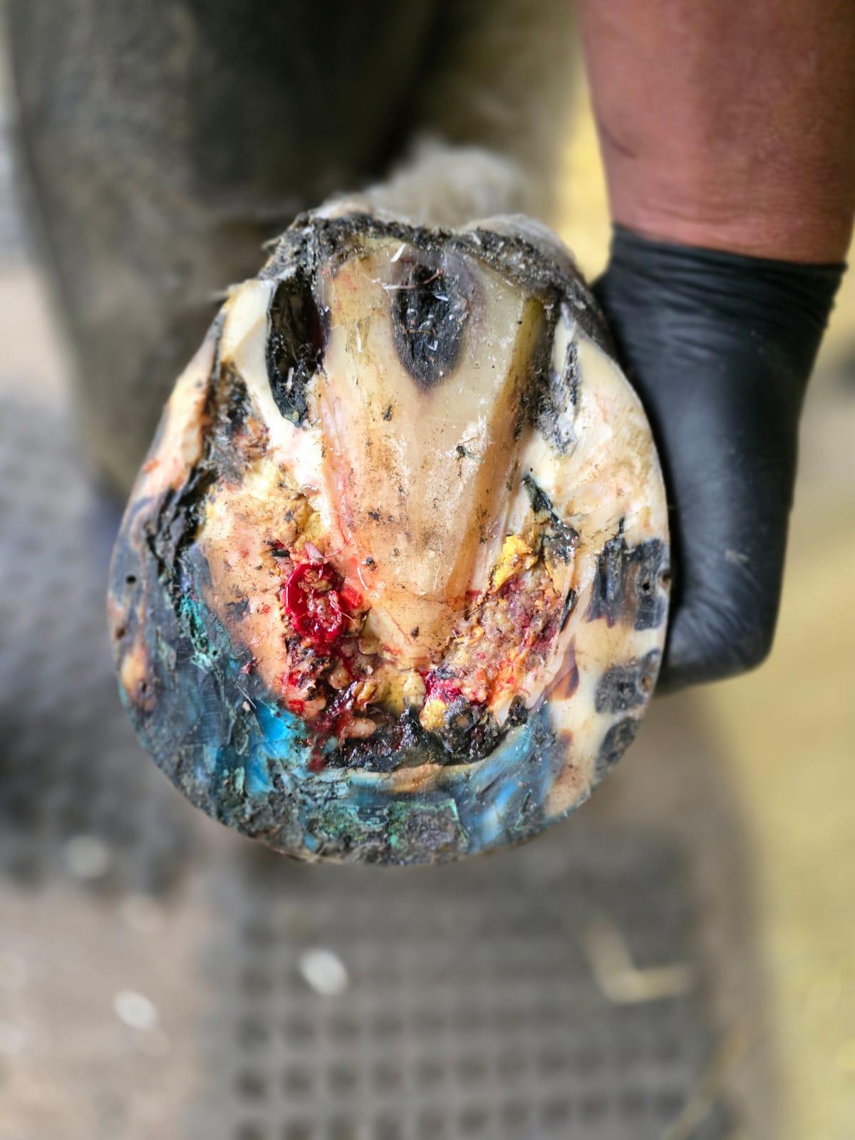
fig 1. severley foundered foot exhibiting gross solear prolapse
Materials and Methods
A modular two-part therapeutic clog was designed, comprising:
- A deformable EVA (ethylene-vinyl acetate) foot-facing surface for improved sole conformity and pressure distribution.
- A rigid external ground surface with integrated options for elevation and graduated palmar angles.
- A removable treatment plate, allowing veterinary access and re-assessment without disturbing the mechanical support.
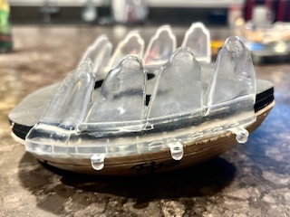
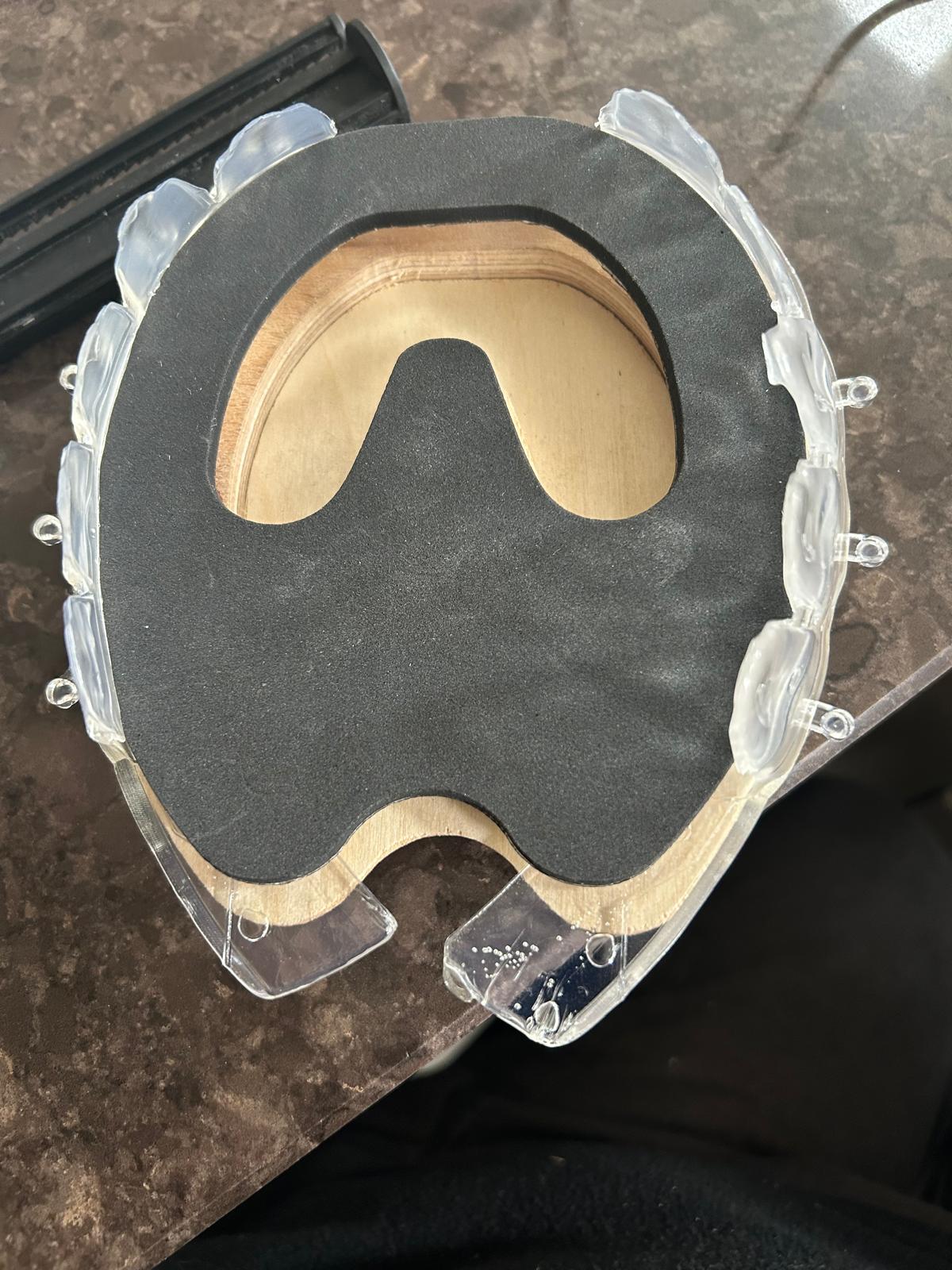
fig 2 the two part glue on hospital plate clog system with EVA rim pad
The clog was applied using a non-invasive glue-on system, avoiding nail trauma in compromised feet.
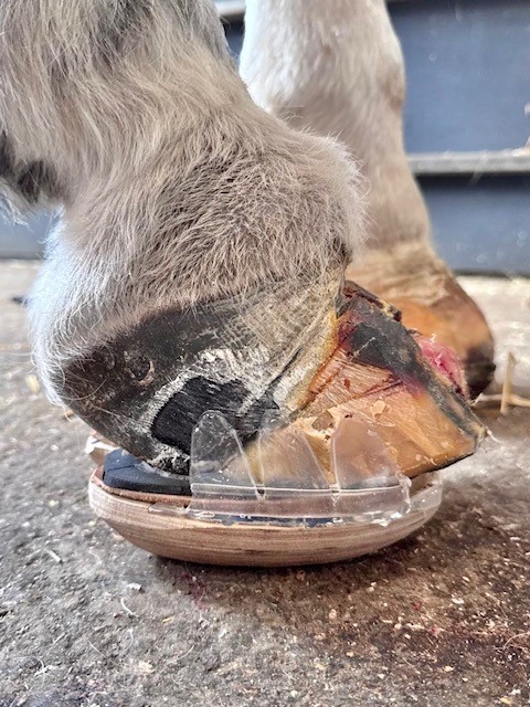
Fig 3. Application of the clog system was non invasive utilising glue on technology (3rd Millenium Co. Ltd)
Data was collected from clinical cases, radiographs, and hoof scanner imaging, assessing:
- Hoof wall lamellar zone width (as per Redden’s protocol).
- Sagittal radiographic measurement between the centre of PIP-DIP articulation and P3 rotational centre.
- Palmar angle variations.
- Clinical outcomes over 8-week follow-ups.
Results
Preliminary data suggests a correlation between increased lamellar zone width and increased sagittal displacement between articulation and rotation centres. Altered palmar angles were also associated with these displacements.
The modular clog system demonstrated:
- Improved load-sharing.
- Reduction in dorsal wall stress.
- Increased palmar support.
- Enhanced patient comfort and mobility.
Radiographic and clinical follow-up showed stabilisation or improvement in P3 positioning in the majority of cases.
Discussion
The study proposes that:
- Lamellar failure and P3 displacement represent both vertical and rotational biomechanical instability.
- Redistribution of ground reaction forces via a modular clog reduces dorsal tension and supports the palmar hoof.
- The EVA foot-facing material offers controlled deformability, increasing surface contact and reducing peak pressure points.
- The removable plate permits ongoing treatment adjustments without disturbing the established load distribution.
- Glue-on application eliminates further wall trauma, crucial in compromised laminitic feet.
These principles are consistent with biomechanical theory and support published findings on heel elevation, positive palmar angles, and deep digital flexor (DDFT) tension reduction.
Conclusion
The modular therapeutic clog system represents a biomechanically sound, adaptable, and clinically effective treatment for equine laminitis. Its design incorporates evidence-based mechanical principles and modern manufacturing processes, with significant potential for improved patient outcomes.
Proposed Future Work
- Pilot studies with quantitative radiographic and scanner data from 50+ cases.
- Development of 3D scanning-based clog customisation.
- Statistical correlation analysis between lamellar zone width, rotational displacement, and palmar angles.
References
Redden, R.F. (2003). Clinical Techniques for Radiographic Analysis of the Equine Foot. Veterinary Clinics of North America: Equine Practice, 19(2), pp. 379-394.
Eustace, R.A. (1992). Laminitis: Current Concepts. Equine Veterinary Education, 21(12), pp. 628–638.
O’Grady, S.E. & Steward, M.L. (2009).Use of Wooden Shoes for the Management of Laminitis. Equine Veterinary Education, 4(2), pp. 61–64.