
A Comprehensive Review of Professor Chris Pollitt’s Contributions to Laminitis Research: Anatomy, Pathophysiology, and Clinical Implications
Caldwell M.N., Madden N,.and Pell C. A review of laminitis research. HoofFlix Scientific.
Key words:
Word count : 5660
Abstract
Laminitis remains one of the most debilitating diseases in the equine population, causing profound pain, loss of function, and in many cases, euthanasia. For decades, it has been misunderstood, with misconceptions regarding its etiology, progression, and management persisting across veterinary, farriery, and equine management communities. Professor Chris Pollitt’s extensive body of work over nearly half a century has fundamentally transformed this understanding. Through meticulous research into lamellar microanatomy, lamellar biomechanics, biochemical cascades, endocrine influences, and diagnostic imaging, Pollitt has provided a comprehensive evidence-based framework for understanding laminitis. This review synthesises the principal themes of his work, explaining how laminitis develops, how structural and biochemical mechanisms interact, and why early recognition and intervention are crucial. Written for veterinary professionals, farriers, equine students, and informed horse owners, this article aims to translate complex scientific findings into accessible, accurate knowledge while preserving technical integrity.
Introduction
Laminitis, the inflammatory and degenerative condition of the equine hoof, represents one of the greatest welfare challenges in the domestic horse population. Clinically, it manifests through pain, lameness, and structural compromise of the hoof, often resulting in catastrophic outcomes if left untreated. Traditionally, laminitis was attributed primarily to vascular compromise or dietary overload, and even today, misconceptions abound regarding its pathophysiology and management.
Professor Chris Pollitt’s research revolutionized this understanding by combining detailed anatomical investigation with experimental and clinical observations. He elucidated the structure of the lamellar interface, clarified the role of biochemical mediators in lamellar failure, and defined the mechanical and endocrine factors contributing to the disease. His work has provided the foundation for evidence-based veterinary and farriery interventions, informed preventative strategies, and established the conceptual framework for understanding both acute and chronic laminitis.
This review follows a narrative approach designed to educate readers with limited prior knowledge of equine anatomy or pathology. It integrates findings from Pollitt’s publications into a cohesive story, highlighting the progression of laminitis from its earliest structural changes to long-term hoof deformation. Key themes include lamellar anatomy, cellular and biochemical mechanisms, endocrinopathic laminitis, mechanical failure, chronic hoof adaptation, diagnostic imaging, and the implications for clinical management and farriery.
Lamellar Anatomy: The Foundation of Structural Integrity
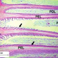
A cornerstone of Pollitt’s contributions lies in his detailed characterization of the lamellar interface of the equine hoof. The distal phalanx (P3) is suspended within the hoof capsule by a complex network of primary and secondary lamellae, collectively forming the suspensory apparatus of the distal phalanx (SADP). Prior to Pollitt’s work, this region was often misrepresented as a relatively simple interface; his studies revealed a highly intricate, interdigitating system optimised for load distribution and structural stability.
Each hoof contains approximately 550–600 primary epidermal lamellae, each of which branches into around 100–150 secondary lamellae. This architecture multiplies the contact surface between the hoof wall and the distal phalanx more than fifty-fold, creating a system capable of suspending the horse’s weight and absorbing substantial mechanical forces during locomotion. The lamellae are anchored by hemidesmosomal attachments to a continuous basement membrane, which itself is intimately associated with the underlying dermal lamellae.
Pollitt’s use of light microscopy, transmission electron microscopy (TEM), and scanning electron microscopy (SEM) allowed him to visualise the lamellae in unprecedented detail. He demonstrated that these structures are not static but are biomechanically dynamic, responding to compressive and tensile forces during locomotion. Furthermore, he highlighted the critical importance of the basement membrane in maintaining structural integrity: its degradation is a primary initiating event in laminitis.
Understanding the lamellar architecture is essential because it provides context for interpreting the earliest changes in laminitis. When the structural interface begins to fail, it is the elongation and detachment of secondary lamellae that herald the onset of disease — often before overt clinical signs such as lameness are visible. This realisation fundamentally shifted the conceptual model of laminitis from one centred on vascular compromise to one emphasising structural and biochemical integrity.
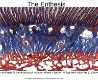
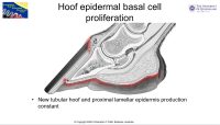
Micro anatomy of the affected primary and secondary lamella in laminitis
Early Structural Changes and Biochemical Mechanisms
One of Pollitt’s most significant discoveries was that lamellar failure begins at the cellular and molecular level long before mechanical collapse is apparent. Through detailed histological studies, he observed that the secondary epidermal lamellae elongate, stretch, and lose their interdigitation with the dermal lamellae early in disease progression. This structural weakening compromises the ability of the hoof capsule to suspend the distal phalanx.
Biochemically, Pollitt identified that matrix metalloproteinases (MMPs) play a crucial role in lamellar degradation. These enzymes degrade components of the extracellular matrix, particularly type IV collagen in the basement membrane. The activation of MMPs can occur in response to systemic inflammatory stimuli, metabolic stress, or endocrine factors. Critically, Pollitt showed that these enzymatic processes precede gross lamellar detachment, providing a mechanistic explanation for the progression of laminitis from subclinical to overt disease.
In addition to enzymatic activity, Pollitt highlighted the importance of cytoskeletal and cellular alterations in keratinocytes. Stretching, apoptosis, and cytoskeletal disruption within the lamellar epidermis contribute to the weakening of the interface. These observations underscore that laminitis is not purely a vascular or inflammatory condition but a disease of structural failure at the cellular level.
Endocrinopathic Laminitis: Hyperinsulinaemia and Metabolic Influence
Pollitt’s work on endocrinopathic laminitis transformed our understanding of the disease’s systemic causes. He demonstrated, through controlled experiments, that prolonged hyperinsulinaemia alone is sufficient to induce laminitis in healthy ponies. This finding explained the prevalence of laminitis in horses with Equine Metabolic Syndrome (EMS) and Pituitary Pars Intermedia Dysfunction (PPID) and challenged the long-standing focus on sepsis or dietary overload as primary causes.
Mechanistically, Pollitt and colleagues showed that insulin interacts with insulin-like growth factor-1 (IGF-1) receptors in the lamellar epidermis, promoting excessive keratinocyte stretching and compromising the mechanical integrity of the lamellae. This endocrine-driven pathway occurs independently of inflammation or vascular obstruction, highlighting a previously unrecognized metabolic etiology. Clinically, this insight has led to preventative strategies focusing on metabolic control, dietary management, and early recognition of at-risk horses.
The recognition of endocrinopathic laminitis as a distinct disease entity underscores Pollitt’s commitment to evidence-based classification. By differentiating between inflammatory, metabolic, and supporting-limb causes of laminitis, practitioners can tailor interventions more effectively, improving welfare outcomes.
Chronic Laminitis and Hoof Capsule Adaptation
Pollitt’s research extended beyond the acute phase of laminitis to the chronic stage, where structural deformities and hoof growth abnormalities become evident. He demonstrated that chronic laminitis involves progressive displacement of the distal phalanx, either through rotation, sinking, or asymmetrical shifts. As the bone moves relative to the hoof capsule, the lamellae adapt or remodel, producing characteristic signs such as divergent growth rings, dorsal wall elongation, and sole flattening.
Importantly, these changes are not purely cosmetic; they reflect ongoing mechanical stress and altered load distribution. Pollitt’s longitudinal observations revealed that chronic laminitis often results in permanent alterations to the basement membrane and dermal structures, emphasizing that early intervention is critical to prevent irreversible damage.
This understanding directly informs farriery practices. Shoe selection, trimming techniques, and weight redistribution strategies must consider the altered biomechanics of the chronic laminitic hoof. Pollitt’s work provides the anatomical and biomechanical rationale for these interventions, bridging veterinary science and practical hoof care.
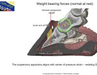
Mechanical Loading and the Suspensory Apparatus
Another key insight from Pollitt’s research is the role of mechanical forces in lamellar failure. The lamellae function as both a suspension system and a shock absorber, distributing weight and mitigating stress on the distal phalanx. When lamellae are weakened, even normal forces during stance or locomotion can exacerbate detachment and rotation.
Pollitt’s studies highlight that the combination of biochemical weakening and mechanical overload is central to disease progression. This mechanistic understanding informs farriery strategies, including heel elevation, toe-offloading, and supportive shoeing, all designed to reduce tensile stress on compromised lamellae.
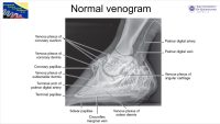
Digital Venography: Evaluating Vascular Integrity
Although laminitis is not initially caused by vascular compromise, Pollitt demonstrated that vascular changes occur as a secondary consequence. Digital venography provides a non-invasive method to visualise perfusion within the foot. Pollitt’s work, along with others Redden et al:, showed that venographic patterns correlate with the extent of lamellar damage and distal phalanx displacement, providing valuable prognostic information.
By differentiating primary lamellar failure from secondary vascular changes, practitioners can avoid misdiagnosis and better plan therapeutic interventions.
Hoof Growth, Horn Production, and Regeneration
Pollitt also contributed to understanding how laminitis affects hoof growth. Damage to the coronary band and lamellar epidermis alters horn production, often leading to poor-quality or distorted hoof wall. These changes are compounded by mechanical stress, resulting in abnormal tubule orientation, sole prolapse, and heel collapse. Recognizing these processes allows farriers and veterinarians to develop corrective strategies that encourage more normal growth and protect compromised structures.
Clinical and Farriery Implications
Pollitt’s work has reshaped the practical management of laminitis. Key implications include:
Early Detection: Recognizing subclinical lamellar stretching and biochemical changes allows for timely intervention.
Metabolic Management: Monitoring and controlling insulin and glucose levels in at-risk horses prevents endocrinopathic laminitis.
Biomechanical Intervention: Corrective shoeing and trimming reduce mechanical stress on the distal phalanx and lamellae.
Long-Term Planning: Chronic cases require an integrated approach addressing structural adaptation, vascular health, and horn growth.
By providing a unifying framework, Pollitt’s research enables collaboration between veterinarians and farriers, ensuring that interventions are both anatomically informed and mechanically appropriate.
Conclusion
Professor Chris Pollitt’s contributions to laminitis research have transformed our understanding of this complex disease. His meticulous work on lamellar microanatomy, biochemical pathways, endocrine influences, mechanical principles, chronic hoof adaptation, and diagnostic imaging has provided a comprehensive, evidence-based model for both prevention and treatment.
For practitioners, farriers, and equine enthusiasts, Pollitt’s research emphasizes that laminitis is a progressive condition of structural failure, influenced by metabolic, mechanical, and biochemical factors. Early recognition, metabolic management, and anatomically-informed interventions are essential to preserving hoof function and equine welfare.
This review synthesises Pollitt’s discoveries into a coherent narrative suitable for educational purposes, providing readers with the knowledge to interpret, prevent, and manage laminitis using current scientific evidence.
For information on the forthcoming webinar series with Professor Pollitt visit
References.
Asplin, K.E., Sillence, M.N., Pollitt, C.C. & McGowan, C.M., 2007. Induction of laminitis by prolonged hyperinsulinaemia in clinically normal ponies. Veterinary Journal, 174, pp.530–535.
de Laat, M.A., McGowan, C.M., Sillence, M.N. & Pollitt, C.C., 2010. Hyperinsulinaemia and lamellar pathology in insulin-infused horses. Equine Veterinary Journal, 42, pp.58–63.
Johnson, P.J., Tyagi, S.C., Katwa, L.C. & Pollitt, C.C., 1998. Matrix metalloproteinases in equine laminitis. American Journal of Veterinary Research, 59, pp.882–887.
Pollitt, C.C., 1992. Basement membrane pathology: a feature of acute equine laminitis. Equine Veterinary Journal, 24, pp.27–34.
Pollitt, C.C., 1996. Basement membrane pathology in equine laminitis: a comparison of two models. Equine Veterinary Journal, 28, pp.38–46.
Pollitt, C.C., 2004. Equine laminitis: a revised pathophysiology. AAEP Proceedings, 50, pp.525–528.
Pollitt, C.C. & Daradka, M., 2001. A cryo-microscope study of lamellar pathology in a horse model of laminitis. Veterinary Pathology, 38, pp.292–301.
Redding, W.R. & Pollitt, C.C., 2010. Equine digital venography: interpretation and clinical application. Equine Veterinary Education, 22, pp.88–95.